• UCLA Health
  • myUCLAhealth
  • School of Medicine
Pediatric Neurosurgery

Pediatric Neurosurgery

Pediatric Neurosurgery
  • About Us
    • Family-Centered Care
    • Chase Child Life
    • In the News
    • Webinars
  • Meet Our Team
  • Conditions & Treatment
    • Pediatric Brain Tumor Program
    • Pediatric Chiari and Syringomyelia Program
    • Pediatric Concussion Program
    • Pediatric Craniosynostosis Program
    • Pediatric Epilepsy Surgery
    • Pediatric Hydrocephalus Program
    • Pediatric Neurovascular Program
    • Pediatric Scoliosis Program
  • Resources for Families
    • A Guide to Preparing for Surgery
    • Admission Information
    • Facilities & Amenities
    • Visitors
    • Medical Records
    • UCLA Neurosurgery App
  • Giving
  • Patient Stories
    • Rhianna's Story - Chiari Malformation
    • Cameron’s Story - Brain Tumor
    • Kobe's Story - Using Virtual Reality for Brain Surgery
    • Brock's Story - Brain Seizures
    • Nathan's Story - Tuberous Sclerosis Complex
    • Nikki's Story - Epilepsy
    • Aiden's Story - Sturge-Weber Syndrome
    • Lucy's Story - Tuberous Sclerosis
    • Nichele - Spinabifida
  • Contact Us
    • Appointment Request
    • Telemedicine and Remote Second Opinion
    • Maps and Directions
  • UCLA Health
  • myUCLAhealth
  • School of Medicine

Pediatric Neurosurgery

Conditions & Treatment

Conditions & Treatment

Conditions & Treatment

  • Pediatric Brain Tumor Program
    • Brain Tumors
    • Our Physician Team
    • Referring Physicians
  • Pediatric Chiari and Syringomyelia Program
    • Faculty
    • Frequently Asked Questions
  • Pediatric Concussion Program
  • Pediatric Craniosynostosis Program
  • Pediatric Epilepsy Surgery
    • Epilepsy FAQs
    • Diseases and Conditions
      • Focal Cortical Dysplasia
      • Hemimegalencephaly
      • Rasmussen’s Encephalitis
      • Sturge-Weber Syndrome
    • Diagnostic Operations
    • Epilepsy Treatment
      • Hemispherectomy
      • Laser Interstitial Thermal Therapy
    • Our Epilepsy Team
    • Patient Services
    • Patient Stories
    • Helpful Links
    • Hemispherectomy Outcome Prediction Scale (HOPS)
    • High Frequency Oscillation
    • Rare Epilepsies & Brain Disease Tissue Bank (REBDTB)
      • Participate in Ongoing Research
      • Donate Biological Samples To The Tissue Bank
      • Submit a Proposal For Research
      • About the Rare Epilepsies & Brain Disease Tissue Bank
      • Contact the Rare Epilepsies & Brain Disease Tissue Bank
  • Pediatric Hydrocephalus Program
    • Hydrocephalus FAQs
    • Faculty
  • Pediatric Neurovascular Program
  • Pediatric Scoliosis Program
  • Pediatric Brain Tumor Program
  • Pediatric Chiari and Syringomyelia Program
  • Pediatric Concussion Program
  • Pediatric Craniosynostosis Program
  • Pediatric Epilepsy Surgery
  • Pediatric Hydrocephalus Program
  • Pediatric Neurovascular Program
  • Pediatric Scoliosis Program
  • Epilepsy FAQs
  • Diseases and Conditions
  • Diagnostic Operations
  • Epilepsy Treatment
  • Our Epilepsy Team
  • Patient Services
  • Patient Stories
  • Helpful Links
  • Hemispherectomy Outcome Prediction Scale (HOPS)
  • High Frequency Oscillation
  • Rare Epilepsies & Brain Disease Tissue Bank (REBDTB)
  • Focal Cortical Dysplasia
  • Hemimegalencephaly
  • Rasmussen’s Encephalitis
  • Sturge-Weber Syndrome
  1. Home
  2. Conditions & Treatment
  3. Pediatric Epilepsy Surgery
  4. Diseases and Conditions
  5. Sturge-Weber Syndrome

Sturge-Weber Syndrome

Share this

Epilepsy Surgery for Sturge-Weber Syndrome

In this section: Tuberous Sclerosis Complex | Focal Cortical Dysplasia | Hemimegalencephaly | Rasmussen’s Encephalitis | Sturge-Weber Syndrome

Sturge-Weber Syndrome at UCLA

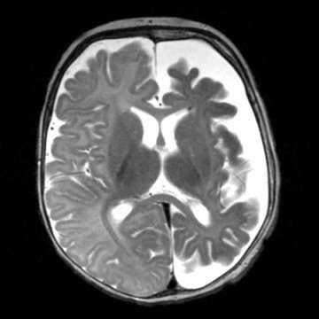
Sturge-Weber syndrome (SWS) is a rare vascular disorder present at birth that is characterized by facial blood vessel malformation (port wine stain) and associated blood vessel malformations affecting the brain and eye. It is not a heritable disorder.  The “port-wine stain” on the face is typically isolated to one half of the face and commonly involves the upper portion of the face.  This skin lesion is usually obvious at birth although its appearance can change with age as the child grows.

Sturge-Weber Syndrome

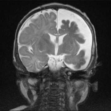
The cause of SWS is a somatic (i.e. non-inheritied) mosaic mutations in the GNAQ gene. The blood vessel malformations associated with SWS can lead to specific neurologic and ocular abnormalities.

Neurologically, the infant or child may present with seizures that are progressively more difficult to control with medications.  In addition, neurological deficits and developmental delay may result.  Visual field defects may also occur with variable severity.   Hydrocephalus (i.e. too much brain fluid) also may occur. A small proportion of patients have no neurologic abnormalities.

The reasons for neurologic progression in SWS are uncertain. One possible mechanism is the lack of blood flow and oxygen to the surrounding brain.

Port wine stain — Cutaneous port wine stain (capillary malformation) is the most common type of vascular malformation, occurring in 0.3 percent of newborn infants (picture 1). However, only a small proportion of children with port wine stains have SWS. (See "Capillary malformations (port wine stains) and associated syndromes".)

In SWS, the port wine stain typically is present on the forehead and upper eyelid, primarily in the distribution of the first or second division of the trigeminal nerve

Sturge-Weber Syndrome

Although traditionally considered important in determining meningeal involvement risk, the location of the capillary malformation (port wine stain) on the face is not a good indicator of the risk of an underlying meningeal capillary malformation. However, the midface and the meninges do share a common embryological origin [4]. Extension of the skin lesion to both sides of the face and the trunk and extremities is common.

The skin lesion usually is obvious at birth. However, its appearance changes with age and its size increases as the patient grows. In the newborn, the lesion is flat and usually light pink in color. It typically darkens with age to a deep red, port wine appearance, and vascular ectasias develop (picture 2). The vascular ectasias produce nodularity and superficial blebbing, which lead to overgrowth of the underlying soft tissues and sometimes the bone. Dental abnormalities may occur as a result of gingival thickening if this area is affected. By the fifth decade, 65 percent of patients with a facial port wine stain have hypertrophy and nodularity within the lesion.


< Return to Diseases and Conditions

Like Us on Facebook Follow Us on Twitter Subscribe to Our Videos on YouTube Follow us on Instagram Connect with Us on LinkedIn Follow us on Pinterest
UCLA Mattel Children’s Hospital rated among best children’s hospitals in U.S.
  • UCLA Health
  • Find a Doctor
  • School of Medicine
  • School of Nursing
  • UCLA Campus
  • Directory
  • Newsroom
  • Subscribe
  • Patient Stories
  • Giving
  • Careers
  • Volunteer
  • International Services
  • Privacy Practices
  • Nondiscrimination
  • Billing
  • Health Plans
  • Emergency
  • Report Broken Links
  • Terms of Use
  • 1-800-UCLA-MD1
  • Maps & Directions
  • Contact Us
  • Your Feedback
  • Report Misconduct
  • Get Social
  • Sitemap
Like Us on Facebook Follow Us on Twitter Subscribe to Our Videos on YouTube Follow us on Instagram Connect with Us on LinkedIn Follow us on Pinterest

Sign in to myUCLAhealth

Learn more about myUCLAhealth