Cellular and Molecular Biology of the Inner Ear Laboratory
Our laboratory is focused on studying the molecular anatomy of the inner (internal) ear in animal models and humans. To study the organization of the internal ear we use a multidisciplinary approach that includes the use of classical anatomical techniques as well as modern cellular and molecular biological methods: immunocytochemistry, transmission electron microscopy, real-time RT-PCR (for gene expression) and, recently, proteomics. We strongly believe that the combination of findings generated with animal and human inner ear research will help us understand its normal and pathological function and provide the basis for new treatments and prevention of hearing loss.
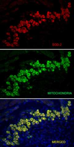
The auditory spiral ganglia, stained with antibodies against the oxidative stress enzyme Superoxide dismutase-2 (SOD-2, red color), and antibodies against itochondria (green color). Merged image at the bottom shows colocalization of SOD-2 and mitochondria. Blue color shows DNA in cell nuclei.
The auditory spiral ganglia, stained with antibodies against the oxidative stress enzyme Superoxide dismutase-2 (SOD-2, red color), and antibodies against itochondria (green color). Merged image at the bottom shows colocalization of SOD-2 and mitochondria. Blue color shows DNA in cell nuclei.
Current Research Projects
Carbon monoxide impairs inner ear development: In this project, we have found that rat pups exposed to mild levels of carbon monoxide (CO) at levels deemed safe by the U.S. government (between 25 and 50 parts per million) suffered hearing and cerebellar dysfunction. CO is an odorless, colorless, poisonous gas that is a byproduct of combustion. In the environment, it is commonly produced by tobacco smoke and auto emissions. Part of this work was published in two peer-review journals: Neuroscience and BMC Neuroscience. In these studies, we analyzed the sensory receptor cells and their neurons in the cochlea, as well as the cerebella of rat pups. Damage to hearing was seen with exposure to as little as 25 parts per million. We found some damage to the neurons in the inner ear of the rats exposed to CO (there was a loss of nerve fibers that send information to the brain). Based on the findings, we believe that infants exposed to low concentrations of CO may suffer hearing damage. Exposure can result from secondhand smoke and other environmental pollution. Because CO is found in the blood and can cross the blood-placenta barrier, damage to an infant's hearing may occur in the womb. Funded by grant 14RT-0043 from the Tobacco-Related Disease Research Program (TRDRP), University of California.
Funding Information
Grant 14RT-0043 from the Tobacco-Related Disease Research Program (TRDRP), University of California.
Members
- Principal Investigator: Ivan A. Lopez, PhD
- Senior Advisor: John Edmond PhD, Mental Retardation Research Center, UCLA
- Post Grad Research Fellows: Luis Beltran Parrazal, PhD
- Undergraduate Students: Amy Ngan, UCLA; Aileen Ngan, UCLA; Yeshika Sharma, UCLA; Tiranun Rungvivatjarus, UCLA; Astkhik Mangansarian, UCLA
- Visiting Students: Francisco A. Mercado, PhD, University of Puebla, Mexico.
Recent Publications
- López IA, Acuna D, Beltran-Parrazal L, Weber D, Espinosa-Jeffrey A, Edmond J.. Oxidative stress and the deleterious consequences to the rat cochlea after prenatal and pre-postnatal chronic mild exposure to carbon monoxide in air. Neuroscience, 151:854-867, 2008.
- López IA, Acuna D, Beltran-Parrazal L, Lopez IE, Cortes M, Amarnani A, Edmond J. Evidence for oxidative stress in the developing cerebellum of the rat after chronic mild carbon monoxide exposure (0.0025% in air). BMC Neuroscience 2009, 10:53, 1-18.
- López IA, Rosenblatt MI, Kim C, Galbraith GG, Jones SM, Kao L, Newman D, Liu W, Yeh S, Pushkin A, Abuladze A, Kurtz I. Slc4a11 gene disruption in mice: Cellular targets of sensorineuronal abnormalities J Biol Chem, 2009, 284:26882-96.
Highlights/News
UCLA Newsroom: UCLA study reveals how tiny levels of carbon monoxide damage fetal brains >
Contact Information
Ivan A. Lopez, PhD:
[email protected]
(310) 825-5331