Radiology
UCLA radiologists combine advanced technology with compassionate, accessible care.


Why choose UCLA Health for radiology?
At UCLA Health, our radiology team combines experience and technology to deliver diagnostic imaging services. We have achieved international recognition for our research programs. Many of our faculty are known worldwide as leaders in their field.
Highlights of the Department of Radiology include:
Full range of diagnostic services: Our radiology specialists provide all types of medical imaging services, both on an inpatient and outpatient basis. We offer specialized services in breast imaging, pediatric imaging, acute care imaging and more. These specializations mean that you receive the most advanced options for your condition and needs.
State-of-the-art technology: Our radiologists use advanced techniques to give you the most accurate radiological diagnosis possible. For example, we have a quantitative three-dimensional imaging lab where we combine high-resolution 3D images with powerful software. This technology gives us unparalleled ability to plan efficient and effective procedures.
Smooth transitions of care: At UCLA Health, we make it easy to coordinate care and share imaging results with your primary physician. Through UCLA Radiology Connections, you can upload images directly to your provider through a secure portal. This service is also helpful when you have had imaging at another institution that you’d like to share with us before your radiology appointment.
Access to clinical trials: The UCLA radiology team includes expert researchers who investigate new approaches to diagnosis and treatment. When you come to UCLA Health, you have access to a range of clinical trials. These studies offer patients more care options, including promising new treatments.
Research and innovation: Our interventional neuroradiologists are leaders in research and innovation. For example, we invented a device called the MERCI. The MERCI was the first approved device for opening brain vessels to treat acute stroke.
Our services
The radiology team supports multiple clinical areas throughout UCLA Health. We provide diagnostic imaging services for breast cancer prevention, varicose vein treatment, lung cancer screening and more. Our services include:
Diagnostic and imaging services
Our radiologists offer a range of diagnostic and imaging services at our radiology centers, including:
Breast imaging: We continually lead the way in breast cancer prevention and diagnosis. Our Mammography Center was the first of its kind in Los Angeles, and we were the first center in the area to offer breast ultrasound. We also were among the first centers in the region with the expertise and resources to offer 3D mammograms, which give us a better view of breast tissue.
Hereditary hemorrhagic telangiectasia (HHT): HHT is a disorder in which blood vessels do not form properly. It primarily affects the skin, lungs, brain or liver. We have achieved certification with the HHT Foundation as a Center of Excellence in HHT treatment. We provide full spectrum, coordinated care for screening, diagnosing and treating HHT.
Inferior Vena Cava (IVC) Filter Clinic: An IVC filter is a small device that prevents blood clots from moving to your heart and lungs. Our interventional radiologists use advanced technology to place, manage and remove these filters.
Lung cancer screening: We work with multiple specialists, including those in thoracic radiology, pulmonary medicine and thoracic surgery to detect and screen for lung cancer. Often, people who have lung cancer don’t know they have the disease until it is severe, and then it is harder to treat. We use an imaging test called low radiation dose computed tomography to detect lung cancer before symptoms appear.
Peripherally inserted central catheters (PICC): PICCs are small, hollow tubes placed through a vein in the arm. You may need a PICC to receive blood transfusions, medications or nutrients. Our radiology specialists conduct a full assessment to determine what type of PICC will best meet your needs and then place PICCs through a short procedure.
Prostate imaging and treatment: We provide comprehensive and sophisticated imaging to detect, diagnose and treat prostate cancer. New technology helps us diagnose prostate cancer sooner and helps us assist surgeons in planning minimally invasive surgery.
Treatments and procedures
Radiologists may also provide treatment for certain conditions. Treatment options we offer include:
Interventional neuroradiology: Interventional neuroradiology is a specialized field that involves using imaging and small plastic tubes to treat brain conditions. We can use these techniques to treat aneurysms, strokes and rare conditions such as Moyamoya disease.
Interventional oncology: This field of oncology focuses on image-guided cancer treatment, including biopsies and shrinking tumors. At UCLA Health, our team pioneered and developed the field of interventional oncology. Now, we perform more than 500 procedures each year, making us one of the largest programs of this kind in the United States.
Interventional radiology: With interventional radiology, we use minimally invasive techniques to treat conditions that used to require open surgery. These techniques lower patient risk and reduce recovery times. We treat complex conditions safely and effectively with minimal scarring or pain.
Neurosurgery treatment for essential tremors: Our radiologists partner with the neurosurgery team to offer an advanced treatment called magnetic resonance guided focused ultrasound. This technique uses a special scanner, helmet and ultrasound waves to treat the part of the brain that causes tremors. It can be an effective treatment for patients with Parkinson’s disease.
Uterine Fibroid Program: We use a unique team approach to diagnose and treat uterine fibroids. Your care team includes the expertise of gynecologists, urogynecologists, interventional radiologists and nurse practitioners. Together, we offer tailored treatment plans that promote women’s long-term health.
Varicose vein treatment: Our radiologists offer comprehensive vein care and treatment for venous insufficiency. Venous insufficiency causes the veins to swell and enlarge, leading to spider veins or varicose veins. We use a patient-centered approach to care where we discuss treatments with the goal of improving your quality of life.
Conditions we treat
Radiologists are crucial members of diagnostic teams. We also offer minimally invasive treatments for a range of conditions, including:
Aneurysms: A bulge or weak spot in one of your arteries, often in the brain or the back of the knee.
Cancers: A large number of diseases where your cells divide uncontrollably, then spread and destroy surrounding tissue.
Fibroids: Noncancerous growths that occur in the uterus, often during a woman’s childbearing years.
Stroke: An interruption to blood supply in your brain, either because of a blockage or a burst blood vessel.
Tumor: An irregular mass of cells that grows uncontrollably.
Tremors: Involuntary contractions or shaking in one part of the body, often in the hands, legs or face.
Varicose veins: Enlarged, twisted veins, most often in the legs.
Tests and procedures radiology centers offer
We use a range of techniques to view what’s going on inside your body. Some of the most common imaging options we use include:
CT: Using a combination of X-rays and a computer to create detailed images of the inside of your body.
Dual-energy X-ray absorptiometry (DEXA): Using small doses of radiation to measure your bone mineral density (the quality and strength of your bones).
Diagnostic ultrasounds: Also called sonography, using high-frequency sound waves to view the structures inside your body.
Fluoroscopy: Using X-rays with a video screen to create a “movie” of the inside of your body.
Mammography: Using low-dose X-rays to examine breast tissue to detect and diagnose breast cancer.
MRI: Using radio waves and magnets to create detailed images of your internal organs and tissue.
Positron emission tomography (PET): A technique that uses a radioactive substance called a tracer to show how your internal organs are functioning.
Meet the UCLA radiology team
Our radiology team is made up of experts in radiological diagnosis and treating a range of conditions. We offer the full range of diagnostic services and use state-of-the-art technology. Our specialists continually research new and promising treatments.
Contact us
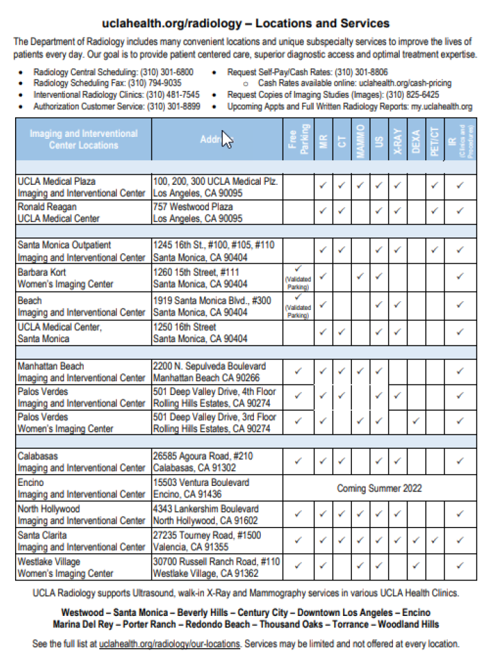
Call 310-301-6800 to request an appointment with a radiologist at UCLA Health or find a medical imaging center near you.
Find your care
Radiologists are experts in all types of imaging, including advanced techniques.
Call today to find medical imaging near you and schedule your imaging procedure (MR, CT, PET, Dexa, Ultrasound).
