Dual Energy CT Angiography
Find your care
Radiologists are experts in all types of imaging, including advanced techniques.
Call today to find medical imaging near you and schedule your imaging procedure (MR, CT, PET, Dexa, Ultrasound).
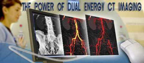
Dual Energy CT Angiography Enhances Diagnostic Capabilities
The dual energy imaging with facilitated bone and plaque removal significantly improves diagnostic confidence of CT angiographic imaging of the entire vascular territory. This technique is particularly helpful in patients with advanced atherosclerosis, when conventional CT angiograms are difficult to interpret.
The Definition Dual Source CT scanner makes non-invasive cardiovascular CT imaging routinely accessible for even the most challenging patients. With a temporal resolution of just 83 milliseconds it can freeze cardiac motion and permits diagnostic imaging of the coronary arteries, pulmonary arteries and aorta at the same time within a single breath-hold. Dual Source CT is not only an excellent diagnostic tool, which improves patient care by making cardiovascular imaging available to even the most critically ill patients, but is also an interesting imaging modality to discover completely new clinical applications.
The Dual Source CT scanner enables the simultaneous operation of the two X-ray sources at two different energy levels (dual energy mode). It therefore allows the acquisition of two data sets of diverse information. Because X-ray absorption is energy-dependent, changing the energy level of the X-ray source results in a material-specific change of attenuation. The material-specific difference in attenuation facilitates differentiation, characterization, isolation, and distinction of the imaged tissue and material. Furthermore, specific details of the scanned body part beyond morphology can be obtained.
The dual energy technique simplifies bone and plaque removal.
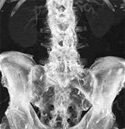
In this context, the dual energy technique has markedly simplified bone removal on cerebrovascular, abdominal and peripheral run-off studies, replacing a laborious and time-consuming manual chore by an easy step that takes less than a minute to perform on a workstation for image analysis and post-processing.
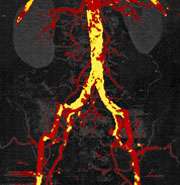
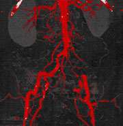
In addition, based on a newly developed software algorithms, the dual energy technique allows the automated removal of calcified plaque from the vessel. This permits to readily detect and display narrowed segments of the arterial tree otherwise obscured by overlying calcifications without time-consuming manual post-processing. This technique is particularly helpful in patients with advanced atherosclerosis, when conventional CT angiograms are difficult to interpret.
Overall, dual energy imaging with facilitated bone and plaque removal overcomes limitations of standard CT, improves diagnostic confidence of CT angiographic imaging of the entire vascular territory and is expected to improve patient care.