Active Surveillance
Find your care
Our radiologists lead the way in prostate imaging. We offer the newest techniques to better detect and stage prostate cancer. Call 310-481-7545 to find out more about prostate imaging and treatment options.
History
- (Initial MRI order was for surgical planning)
- 65 year-old
- PSA 3 → 11 over 6 months
- Microfocus of Gleason 3+3
- Initial report : No posterior disease
- Targeted Biopsy consult prompted 2nd look
Imaging
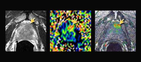
LEFT: Axial T2-weighted image shows slightly asymmetric low signal in the anterior base (yellow arrow)
CENTER: Colorized apparent diffusion coefficient (ADC) map: moderately restricted diffusion
RIGHT: Colorized dynamic contrast enhanced (DCE) perfusion map: focal intense enhancement
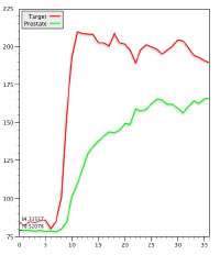
Perfusion time-intensity curve: type III (washout) curve is highly suspicious
Example Report Excerpt for Biopsy Flanning
* Findings
Prostate measures 45 gm with mild prostatic hyperplasia. A single suspicious area is identified:
| Location | Left anterior base 1:00 |
| Size | 1.1 cm |
| Capsule | May involve anterior capsule |
| T2 | Asymmetric, ill-defined (4/5) |
| Diffusion | ADC 0.86, highly restricted (4/5) |
| Perfusion | Intense early + washout (5/5) |
| Overall Suspicion | High (4/5) |
Seminal vesicles and neurovascular bundles appear normal
Results
- Gleason 4+4
- Bone scan: no metatatic disease
Advantage: UCLA Prostate MRI
- MRI in active surveillance can either provide reassurance that no significant disease is missed, or find suspicious areas (usually anterior) that warrant tissue sampling.
- K-space sharing dynamic acquisition (in this case, Siemens TWIST) allows for high spatial and temporal resolution in dynamic contrast-enhanced imaging.