Small Volume Low Grade Disease
Find your care
Our radiologists lead the way in prostate imaging. We offer the newest techniques to better detect and stage prostate cancer. Call 310-481-7545 to find out more about prostate imaging and treatment options.
History
- 60 year-old
- PSA 3.6
- 5 of 6 right biopsies GS3+3
Imaging
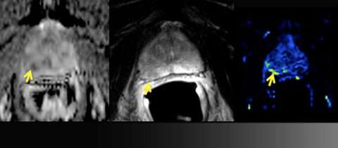
LEFT: Apparent diffusion coefficient (ADC) map: moderately restricted diffusion
CENTER: T2 Weighted imaging: ill-defined low signal (yellow arrow) does not abut the prostate capsule
RIGHT: Perfusion map: focally increased perfusion
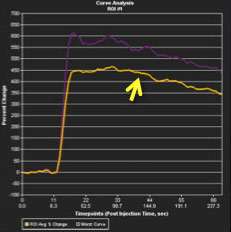
LEFT: Perfusion time-intensity curve: type III (washout) curve is highly suspicious
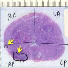
RIGHT: Whole-mount pathology: two low-grade (Gleason Score 3+3=6) tumors do not involve capsule
Advantage: UCLA Prostate MRI
- Although less sensitive for low-grade disease, MRI can help with determining extent and determination whether surveillance or definitive treatment is warranted.
- Diffusion weighted imaging (such as Siemens REVEAL) with multiple b-values allows for high contrast-to-background on ADC map and computation of high b-value images