SOMATOM Definition
Find your care
Radiologists are experts in all types of imaging, including advanced techniques.
Call today to find medical imaging near you and schedule your imaging procedure (MR, CT, PET, Dexa, Ultrasound).
SOMATOM Definition: Faster Than Every Beating Heart
Dual Source Computed Tomography Scanner
UCLA Medical Plaza 200 facility has installed the first Dual Source computed tomography (CT) scanner, SOMATOM Definition, on the west coast. It is the world's first Dual Source CT, using two X-ray sources and two detector arrays at the same time.
The two X-ray sources at right angle to each other make the SOMATOM Definition the fastest CT scanner in the world. With a single source CT scanner, the X-ray source/detector system has to obtain data projections of 180° to take a cardiac image. With the Dual Source CT, each of the two source/ detector combinations only needs to travel 90°, hence double the speed, to acquire diagnostic images of the heart.
Patients with fast, irregular heart rates no longer need beta- blockers. Unlike most other organs, the heart can be regarded as a moving target. Just like in photo- graphy, fast shutter speeds are needed to capture a speeding car free of motion blur. Considering the rapid motion of the heart at about 70-75 beats per minute (bpm) when resting, a single source CT scanner, even a 64-slice CT scanner, is still too slow to visualize the heart without motion artifacts. As a result, beta-blockers have been given to patients with heartbeats greater than 60-65 bpm to slow down their heart rates. With the Dual Source CT, consistent, sharp cardiac images can be acquired independent from the heart rate. Therefore, patients with fast, irregular heart rates or arrhythmia seeking CT exams no longer need beta-blockers.
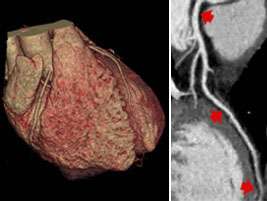
Images acquired from a 46-year-old patient with a heart rate of 78 bpm and no administration of beta blockers.
Left: Volume rendered display of the heart with diagnostic image quality for heart and coronary arteries.
Right: Curved reformatted image of the left anterior descending artery displays coronary artery with high detail information including visualization of small side branches.
"The Technology provided with the new Dual Source CT scanner, the fastest system on the market, allows us to image the beating heart with unprecedented spatial and temporal resolution. And with the integrated 'Care Dose 4D' tool, we are even able to reduce the radiation exposure of the patient."
Stefan Ruehm, MD, PhD
Director of Cardiovascular CT
Professor of Radiology
In addition to its speed, the Dual Source CT offers the ability to better characterize soft tissues. Because X-ray absorption is energy-dependent, changing the energy level of the X-ray source results in a material-specific change of attenuation. With two X-ray sources scanning at different energy levels at the same time, the Dual Source CT scanner acquires two data sets with different attenuation levels simultaneously. The material-specific difference in attenuation may facilitate classification of different tissue types and may help in the characterization and differentiation of different types of atherosclerotic plaque, e.g. calcified & non-calcified plaque. This is expected to improve risk stratification of cardiovascular patients.