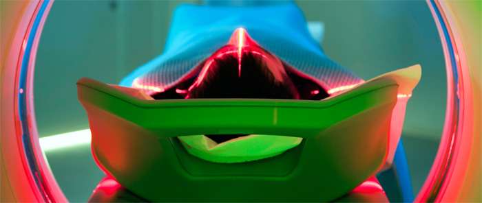
Imaging for Cancer Surgery
Find your care
Our Head and Neck Cancer Program helps you start on the road to recovery. For more information, connect with a cancer care specialist at 310-774-6275.
Expert Head and Neck Cancer Imaging at UCLA
Complete tumor removal is critical in curing head and neck cancer. If any cancerous cells are left behind, many of these cancers may recur. At the UCLA Head and Neck Cancer Program (HNCP), our surgeons use preoperative imaging to assess the size and locations of tumors. Accurate imaging helps ensure that they remove all the cancerous tissue.
Preoperative imaging is used to assess tumor size and location.
The ultimate goal of cancer tumor surgery is “clear margins,” a complete removal of the tumor. Our surgeons are highly skilled in achieving clear margins using imaging and their own training and experience.
- Determining the margin between the healthy and cancerous tissue relies on what the surgeon sees and feels in the affected area during surgery.
- Your surgeon assesses whether the cancer has been removed by taking samples from the surgical margins using frozen section microscopy.
- Your surgeon will make the decision to stop or continue removing tissue based on the preoperative imaging studies, observations of the affected area and the results of frozen section pathology.
Noninvasive Tumor Imaging at UCLA
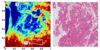
Surgeons balance the goal of achieving clear margins with minimizing the risk of damage to nearby healthy tissue. This concern can limit the amount of tissue that may be safely removed.
To overcome this problem, our surgeons have developed a novel, noninvasive imaging system that can distinguish between cancerous and normal tissue during surgery. Unique to UCLA, this intraoperative instrument allows us to more accurately determine true cancer margins.
We have engineered this system to clearly identify tumor tissue and normal tissue. This tool helps our surgeons remove as much of the tumor as possible without damaging healthy tissue, which improves our patients’ outcomes.
Preoperative Imaging Appointments
- Contact Us >
- Phone: (310) 267-7076