Dural Arteriovenous Fistula (DAVF)
Find your care
Our expert team is skilled at treating complex cerebrovascular conditions and providing the fines and most comprehensive patient care. For help finding a neurosurgeon, call 310-825-5111.
Overview
What is a dural arteriovenous fistula (DAVF)?
Arteriovenous fistulas occur when abnormal connections form directly from arteries to veins. In the nervous system, this direct connection most commonly occurs in the covering of the brain and spinal cord called the dura mater. When this connection occurs in the dura mater, they are called Dural Arteriovenous Fistulas (DAVFs). These fistulas lead to a buildup of pressure in the veins surrounding the brain or spinal cord, and can result in nervous system dysfunction, seizure, and hemorrhage.
Carotid-cavernous arteriovenous fistulas (CCF) are a common subtype of DAVF that occur in the dura and veins behind the eyes. The abnormal connections involve branches of the carotid arteries and the cavernous sinus.
Each dural arteriovenous fistulas is unique and should be evaluated by neurovascular experts, such as the multidisciplinary team at UCLA.
Signs and Symptoms
What are the symptoms of an arteriovenous fistula?
The symptoms of a DAVF can vary widely depending on its size, location, and complexity. Patients with a DAVF sometimes report hearing their own a heartbeat or a whooshing noise in one or both ears, called pulsatile tinnitus. Their physician can sometimes hear this whooshing noise with a stethoscope, and this is called a bruit. Those with CCFs may experience redness and swelling in one or both eyes, as well as double vision. Some common symptoms include:
- Headache
- Pulsatile tinnitus or bruit – whooshing or ringing sound in one ear
- Seizures
- Eye or visual problems – Redness of the eye(s), swelling, double vision are symptoms of CCFs.
- Cognitive decline – In rare cases, longstanding untreated fistulas can lead to brain dysfunction and dementia-like symptoms.
- Stroke-like symptoms
When to see a doctor urgently
Seek urgent medical attention if you notice any signs or symptoms of a stroke, even if they come and go or disappear.
To remember the signs of a stroke, think "FAST" and do the following:
- Face. Ask the person to smile. Does one side of the face droop?
- Arms. Ask the person to raise both arms. Does one arm drift downward? Or is one arm unable to rise? If so, call 911.
- Speech. Ask the person to repeat a simple phrase. Is his or her speech slurred or strange? If so, call 911.
- Time. If you observe any of these signs, call 911 or emergency medical help immediately as time is critical.
Call 911 or your local emergency number immediately. Don't wait to see if symptoms go away. The longer the time before stroke treatment, the greater the chance of brain damage and disability.
Diagnosis
How is an arteriovenous fistula diagnosed?
If a DAVF is suspected, it is important to make a diagnosis and consult with a specialist quickly to prevent worsening of the disease. At UCLA, we use the most advanced technology and latest imaging systems to diagnose arteriovenous fistulas of the brain and spine quickly and accurately. In many cases, a DAVF is discovered once the abnormal blood flow has put enough pressure on the brain to cause symptoms, but whenever possible we try to locate and treat them before severe symptoms result. During your examination, your expert team may use the following imaging techniques to locate and assess your AVF:
- Magnetic resonance imaging (MRI) scans, including magnetic resonance angiography (MRA) and venography (MRV), are used to identify and locate an AVF.
- Catheter angiography provides the most accurate images of the blood vessels affected by the AVF. The help to define the arteries bring blood into the fistula, the regions of abnormal connection to veins, and the pathway of the blood leaving the fistula in the veins. These studies are also used to help plan treatment.
Classification
Dural arteriovenous fistulas (DAVFs) are classified based on the connections between the arteries and veins on the DSA Angiogram and the pathway at which the high-pressure blood flow exits the brain or spine. There are two commonly used grading systems:
The higher the DAVF grade, the higher the risk of brain bleeding or neurologic problems. The higher the DAVF grade, the more likely your cerebrovascular neurosurgeon will recommend treatment. Spinal DAVFs have their own unique symptoms, diagnosis, and treatment considerations. These are also disease entities that often require treatment.
Treatment
How is an arteriovenous fistula treated?
After your diagnosis, a multidisciplinary team of neurovascular experts will meet to review your case and create a customized treatment plan. At UCLA, we typically treat AVFs using strategies that include some combination of:
- Observation: Some fistulas are not immediately dangerous and can be monitored for growth and change without surgery.
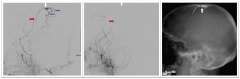
- Endovascular liquid embolization: This minimally invasive technique is performed by inserting a catheter through the groin (or other vessel), then injecting a liquid that solidifies in the blood vessel and cuts off blood flow to the fistula. The cerebrovascular team at UCLA is actively involved in research and testing of new liquid embolic treatments (Figures 1-2). This is the most common method of treating DAVF, and it is often curative.
- Coil embolization: Micro-coils made of platinum are placed into the artery, vein or venous sinus to seal off the DAVF abnormal connection and prevent blood flow through it.
- Microsurgical obliteration: During this open surgery procedure, the neurosurgeon disrupts or disconnects the arteriovenous fistula from the brain or spinal cord. We use high resolution advanced imaging before surgery to map out a patient's precise anatomy and advanced imaging during surgery to guide the procedure.
Postoperative care and recovery
DAVF diagnosis and treatment procedures are performed at Ronald Reagan UCLA Medical Center. Since the procedures are performed using cutting edge technologies and often use minimally invasive technique, there is reduced discomfort and a shortened hospital stay compared to traditional treatments. After treatment, patients are monitored overnight in the hospital and may be discharged as early as the following day.
At UCLA Health neurosciences, we employ highly specialized multidisciplinary health care teams, and in addition to innovative cutting-edge care, we provide support services for patients and their families to help navigate their time in our clinics and hospitals.
Contact us
To schedule an appointment at the UCLA Cerebrovascular Program, please call 310-825-5111 or request an appointment.
Geoffrey Colby, MD, PhD
Jeremiah Johnson, MD