Lung Cancer – Brachytherapy
Find your care
Radiation oncologists use a patient-centered approach and individualized treatment plans. To learn more and make an appointment with a UCLA radiation oncologist, contact us at:
Westwood - 310-825-9775
Santa Clarita - 661-287-0010
Santa Monica - 424-259-8777
El Segundo - 310-794-7301
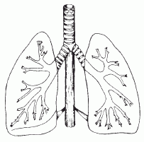
High Dose Rate brachytherapy is used to give an added radiation dose to lung cancers that are located in the major bronchi ( the breathing passages). While increasing the dose to the tumor, HDR, due to its short treatment distance, minimizes the radiation dose given to sensitive nearby tissues such as adjacent normal lung, heart, esophagus and the spinal cord. HDR brachytherapy is also used to treat recurrent endobronchial tumors where additional surgery and external beam radiation therapy are no longer options. "In cases of the tumor being in the lung tissue, rather than the bronchi, external beam radiation therapy, not HDR, is the appropriate therapy."
2. Surgery and HDR Procedure
The patient is made comfortable on the treatment table with intravenous conscious sedation that is typically used in standard bronchoscopic procedures. Topical anesthetic is applied to the back of the nose and throat. An instrument called a bronchoscope is used to locate and evaluate the extent of the tumor. A bronchoscope is a flexible tube that contains a hollow channel and a fiber-optic camera. The pulmonologist inserts the bronchoscope through the nose, past the vocal cords and into the affected bronchus. When the bronchoscope is in position near the tumor, a thin catheter that the radioactive source will travel through is inserted into the bronchoscope. The pulmonologist can see the catheter as it emerges from the end of the bronchoscope. The bronchoscope is then removed, leaving the catheter in place. Usually two catheters are inserted. Special x-ray films are taken in order evaluate the catheter placement and do the computerized treatment plan. When the plan has been approved by the physician, the protruding ends of the catheters are attached to the HDR afterloader, which directs the radioactive source to the precise location in the catheters to deliver the prescribed radiation dose.
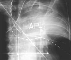
AP (front) radiograph of three treatment catheters placed in the bronchi to encompass the lesion that is obstructing air from reaching the left upper lobe of this patient. After the treatment, the catheters are removed, the patient recovers quickly and then goes home. Usually, three or four treatments are given. Successful treatment opens the bronchi, (improving breathing), and reduces bleeding (hemoptysis).
Our Lung Cancer Experts: