Acoustic Neuroma Program
Find Your Care
Put your trust in our internationally recognized brain tumor experts. For more information, connect with a cancer care specialist at 310-825-5111.
The UCLA Acoustic Neuroma Program
Welcome to the Acoustic Neuroma Program at UCLA
We appreciate your interest in the acoustic neuroma program at UCLA. Under the highly experienced expert leadership of Gregory Lekovic MD, PhD, and Akira Ishiyama MD, we provide comprehensive treatment options for patients with acoustic neuroma/vestibular schwannoma. In addition to providing state-of-the-art care to our patients, we are committed to advancing the field through technical innovation in hearing preservation and rehabilitation, and are leaders in research into the basic science of vestibular schwannoma biology.
Our surgical patients are cared for at the main UCLA campus in Westwood at the UCLA Ronald Reagan Medical Center, consistently ranked one of the best hospitals in the nation, and where anesthesia, radiology, pathology and any other services you might need during your hospital stay are provided by physicians who are leaders in their respective fields.
We are grateful to those who have entrusted us with their care and who have shared their patient experience.
Expert Leadership
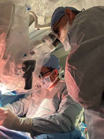
Dr. Gregory Lekovic and Dr. Akira Ishiyama are Co-Directors of the Acoustic Neuroma and Lateral Skull Base Surgery Program at UCLA. They are among the most highly experienced acoustic neuroma surgeons in the country, having performed over 1,500 acoustic neuroma surgeries. While a high volume surgical practice is key to providing the best surgical outcomes, most patients seen by Dr. Ishiyama and Dr. Lekovic do not require active intervention (i.e., observation, aka wait and scan) and many that do need treatment are recommended to undergo stereotactic radiosurgery in lieu of traditional microsurgery.
Their treatment philosophy is truly patient-centered with the most important consideration for most patients being preservation of normal facial nerve function, and whenever possible saving a patient’s natural hearing. That having been said, for patients having surgery, removing the whole tumor is usually the goal unless doing so would jeopardize the facial nerve.
Dr. Lekovic and Dr. Ishiyama have assembled a "dream team" of physicians (including oto-neurology, radiation oncology, neuro-anesthesia, neuro-radiology, neuro-pathology) and allied health professionals (audiology, physical therapy, speech and language pathology) at Ronald Reagan UCLA Medical Center — all dedicated to providing the best possible care for patients with acoustic neuroma.
Comprehensive Treatment Options
At UCLA, our acoustic neuroma program is full of innovation, improving visualization of the tumor in the surgical field and removing tumors atraumatically using new surgical instrumentation, fluorescence visualization.
- Observation, aka “wait and scan”
- We typically get a follow-up MRI six months after the first MRI in which the diagnosis of VS was made. Subsequent MRIs are obtained annually.
- Microsurgery
- There are three main surgical corridors (“approaches”) to acoustic neuroma: the translabyrinthine (aka "through the ear"), retrosigmoid (aka "behind the ear") and middle fossa (aka "above the ear"). At UCLA we have extensive experience in expanded translabyrinthine approaches, which are uncommonly performed but critical for giant and/or medial tumors. We also are pioneers in hybrid approaches combining surgical tumor removal and hearing rehabilitation (e.g., cochlear implantation).
- Radiosurgery
- Stereotactic radiosurgery may be delivered in one, three or five “fractions” depending on the size of tumor and the ability to meet dose constraints (ie dose to the cochlea). We collaborate with radiation oncology to provide you with the safest and least toxic radiation therapy treatment options.
Which treatment is best for you depends generally on three factors: the size of the tumor, your age, and the status of your hearing. In general, the older the patient the more likely it is that radiation will be the treatment of choice, all other things being equal. Youth and large tumors push to surgical management. Hearing can only be preserved for the long term when surgery is done when tumors are small. A current area of investigation is the role of cochlear implants in regaining or rehabilitating hearing when it is either too poor to be considered for a conventional hearing preservation approach or when hearing is lost during attempted hearing preservation surgery.
Innovation
At UCLA, our acoustic neuroma program is full of innovation, improving visualization of the tumor in the surgical field and removing tumors atraumatically using new surgical instrumentation, fluorescence visualization.
- Intra-operative cochleography and cochlear neurography
- Hearing rehabilitation (CI program)
- Novel surgical technique development for CI
- Early facial nerve rehabilitation
- Intra-operative fluorescence visualization of the facial nerve
Research
At UCLA, our acoustic neuroma research program focuses on understanding the basic biology that drives growth of vestibular schwannoma and the factors that cause hearing loss.
- Basic Science
- Temporal Bone Repository
- Surgical Anatomy Laboratory
- Advance imaging initiative
- Mechanism of hearing loss in VS
- Biomarkers of tumor behavior in VS
- Clinical Investigations
- Tissue bank
- Imaging correlates of hearing loss and vestibulopathy in VS
Message from Dr. Gregory Lekovic and Dr. Akira Ishiyama
"At UCLA, we understand that you may have many questions regarding your acoustic neuroma, including what to do about it and when! But the diagnosis of acoustic neuroma is not one that should be feared.
Our mission is to minimize the impact of this diagnosis on your life — for many patients with minimal symptoms, this might mean getting an MRI once a year. For those who need to have surgery we focus on making the experience as safe as possible, with the lowest risk of injury to the facial nerve and whenever appropriate trying to save hearing. We are proud of our superb outcomes but we always strive to do even better!
We would be happy to discuss your case with you, and let you know how your tumor would be treated at UCLA, whether or not you are able to come to Los Angeles for treatment."
What You Will Need
Name, DOB and contact number
MRI/CT and any other imaging
Hearing test results (if available)
Frequently Asked Questions
What is acoustic neuroma/ vestibular schwannoma?
Acoustic neuroma and vestibular schwannoma are synonyms. They are benign (i.e., non-cancerous) tumors that are typically slow growing. They most commonly present with hearing loss, and that is why historically they were referred to as "acoustic" tumors. We now know that they actually arise from the insulating cells (Schwann cells) of the vestibular nerve, hence the more accurate term "vestibular schwannoma."
What are the treatment options?
It is said that there are three treatment options for acoustic neuromas: observation (also called "wait and scan"), surgery, and radiation. Specific treatment recommendations are formulated on an individual basis, and depend on three important variables: tumor size, patient age, and hearing status.
How urgent is treatment of acoustic neuroma tumors?
We prefer not to treat tumors when they are first diagnosed since some of them will remain dormant for years (particularly those diagnosed incidentally) and there is little risk of tumors progressing rapidly. Ideally, we reserve treatment for tumors that are growing or symptomatic. That having been said, tumors sometimes present when very large and treatment in those cases is urgent or even emergent.
What are your outcomes for hearing preservation?
For patients who are ideal candidates for hearing preservation surgery (middle fossa craniotomy), good or useful hearing is preserved approximately two-thirds of the time (meaning AAO-HNS class A or B hearing). It is common for patients to experience a decrement of 10 to 15 dB in hearing after surgery so the better your hearing at the time of surgery, the better the odds of keeping excellent hearing after surgery. The best odds for removing the whole tumor and saving hearing is for small tumors, generally less than 15mm in maximum dimension. Other factors such as the surgical approach and tumor location can also influence the odds for hearing preservation.
Radiosurgery is, like wait and scan, not a hearing preservation treatment strategy.
Will I go deaf or experience hearing loss in the ear with the tumor?
Acoustic neuromas cause hearing loss over time. Hearing loss does not require growth of the tumor, and is at most very loosely correlated with tumor size. Interestingly, the reason these tumors cause hearing loss is not fully understood. Radiation treatment is not compatible with hearing preservation, though whether this is because radiation does not remove the tumor or because of the toxicity of treatment is also unknown.
Therefore, the best way to keep your natural hearing for the remainder of your life is with successful surgery for hearing preservation, assuming the tumor is small enough to be a candidate. Unfortunately, with surgery there is also a significant chance to lose hearing, so surgery is at the same time the best chance to save your natural hearing until old age and simultaneously the quickest way to lose hearing.
There is no risk to the unaffected ear, such that when deafness occurs, it is single-sided deafness only.
If I experience hearing loss, can I get my hearing back?
Generally speaking, hearing does not improve after surgery for tumor removal. In recent years the morbidity of single sided deafness has been better understood and this has led to an increased effort to rehabilitate hearing when lost, whether with a wearable hearing aid or an implanted device such as a cochlear implant or bone anchored hearing aid. At UCLA, we are committed to rehabilitating hearing for every patient, whether deafness occurred before or after treatment.
What are the risks of surgery?
There are three principal risks of surgery: facial nerve injury, CSF leak, and wound infection. The risk of injury to the facial nerve depends on several factors such as tumor size, surgical approach, and prior treatment history (ie prior radiation), but in general is <5%. Cerebrospinal fluid (CSF) can leak through the temporal bone to the Eustachian tube and from there out the nose. We routinely take a piece of fat from the abdomen through a separate incision and use it to plug the hole in the dura through which the CSF enters the wound, but even so there is ~5% risk of CSF leak following surgery. Although the CSF in and of itself is harmless, a leak poses a risk of infection; hence CSF leaks are important to recognize and treat promptly. This typically requires a return to the operating room or the placement of a drain to divert CSF. CSF leaks usually present themselves during the hospital stay, though can occur in delayed fashion.
Wound infections, on the other hand, occur ~1% of the time and typically do not manifest until 7 to 10 days after surgery. Most wound infections are superficial and if caught early do not pose a major risk, though if they are allowed to go deep they may result in meningitis and for this reason wound infections are also treated promptly when recognized. This may require a return to the operating room, though many superficial infections can be managed with oral antibiotics alone.
Fortunately, surgery is very safe, with the risk of major stroke, cardiac arrest, neurological disability or death at 1:1000 or less. Hemorrhages (bleeding) into or around the surgical site may occur as often as 3 to 5% of the time but does not usually require any intervention.
What is stereotactic radiosurgery?
For benign tumors like acoustic neuroma a radiation technique called radiosurgery is preferred. Radiosurgery basically means the delivery of radiation with "surgical intent," i.e., to target the tumor itself and spare the surrounding tissue, as though with a scalpel. The goal of radiosurgery is called "tumor control" by which we mean that the tumor has ceased growth. The goal is not to kill the tumor or melt it away.
Radiosurgery was first introduced for the treatment of acoustic neuroma around 1990 with the Gamma Knife; initially the doses used caused a high risk of facial palsy and so over decades the dose has been refined to the minimal dose that can be given to achieve tumor control- about 12.5 Gy if delivered in a single session. There are many machines that are capable of performing radiosurgery, and sometimes the treatment may be spread over three to five days rather than a single session (called fractions). There are unfortunately very few high-quality studies that compare radiation treatment approaches head-to-head, but if you are being treated with radiation for acoustic neuroma, you should make sure that you are being treated with a radiosurgery treatment plan.
Irrespective of the platform used to deliver radiation, certain commonalities exist. First and foremost is that radiosurgery for acoustic neuroma is very different than radiation delivered for cancer — the dose prescribed is not a lethal dose, but rather a dose designed to change the behavior of the tumor (to prevent growth). If it were a high dose, the collateral damage radiation would do to the facial nerve and brainstem would make the treatment worse than the disease, and the risks of treatment much higher than with surgery. Because the surrounding brain tissue receives so much less radiation with radiosurgery than with conventional radiation, a lot of the side effects one associates with radiation (hair loss, fatigue, nausea, etc.), generally speaking, do not apply to radiosurgery. That having been said, because of the accuracy of radiosurgery and the steep drop-off of dose that these machines are able to achieve, the tumor receives a relatively large dose per fraction. This — relatively high dose to the tumor, low dose to neighboring brain — is the key to obtaining tumor control and minimizing complications.
Finally, following radiosurgery shrinkage of the tumor is not uncommon but is only seen ~40% of the time. Moreover, the effect of radiosurgery treatment is not immediate. It generally take a minimum of six months to halt growth and may take up to two years, so we generally are very reluctant to consider a radiation treatment to have failed before waiting this long. For this reason also, radiation may not be the treatment of choice for tumors that are causing a lot of symptoms by putting pressure on the brainstem or nerves.
Although it has been in use for decades, we still do not fully understand how or why radiation delivered in this way works!
What are the risks of radiation treatment?
The risks, or complications, of radiation treatment can be segregated into three distinct time periods. Early complications following radiation treatment occur in the days during or immediately following completion of treatment and are typically limited to headache, nausea, and are usually well controlled with short course of steroids. Intermediate complications occur usually within six months to two years after treatment, and include facial weakness in 1 to 2% of patients, numbness in 3 to 4%, and adverse radiation effect on the brainstem, also 3 to 4% of the time (but only when the tumor is in contact with the brainstem). The delayed risks of radiation include the induction of new tumors (meningiomas in particular, but also the risk of malignancy) and typically does not occur until 7 or more years after radiation treatment is completed.
In addition, because the tumors are not killed by the radiation treatment, some tumors recover from the effect of radiation and resume growth after a period of time. The radiation failure rate is very low during the five years following treatment (~5%) but may be as high as 15 to 20% at 10 years.
What does recovery from surgery look like?
The recovery from surgery occurs in three stages: in the hospital, after discharge but before the wound has fully healed, and after suture removal (at two weeks post op). After surgery you can expect to spend 3 to 4 nights in the hospital, depending on how dizzy or off-balance you feel. It is very common to be dizzy after acoustic neuroma surgery and recovery of your balance is important prior to discharge. That having been said, you may still feel a little off balance (akin to just disembarking from a boat) for the week or two following surgery.
For the following ten days or so after discharge (ie when at home but before the sutures have been removed), we encourage you to be as active as you can tolerate, especially walking. You will not need any special nursing care or help with your activities of daily living, though it is probably prudent not to be alone.
Until the sutures are removed we ask that you avoid straining (to minimize the risk of CSF leak), so do not lift anything heavier than ten pounds, take a stool softener, and avoid bending over at the waist.
How do I care for my surgical wound?
Wash the wound and shampoo your hair daily after discharge from the hospital. Sutures will be removed approximately two weeks after the day of surgery.
Is radiation or surgery a better treatment option for me?
In comparing radiation and surgery, it is important to understand that aside from the risk/benefit ratio calculation, there is a very fundamental difference in the goals of treatment between radiation and surgery. Specifically, the goal of radiation is to "control" the tumor (i.e., prevent growth) whereas the goal of surgery is to remove the tumor. Because radiation treatments do not kill the tumor, and because hearing loss does not depend on tumor growth, the tumor continues to adversely affect hearing after radiation treatment. So for patients with excellent hearing for whom hearing preservation is technically feasible, consideration may be given to surgery rather than radiation. In addition, some tumors recover from the radiation treatment and resume growth- sometimes years after radiation has been given. This is called radiation treatment "failure." The rates of radiation treatment failure are low, but as high as 15 to 20% at ten years. So for older patients radiation tends to be a safer bet than for younger patients, all things being equal.
The main advantage of radiation is that it is less likely to cause facial weakness compared to surgery. In most published series of radiation treatment, the risk of facial nerve injury is as low as 1 to 2% and as high as 3 to 4%, though in our experience facial weakness after radiation is seldom a complete facial paralysis. In experienced hands, the risks of permanent facial weakness following surgery is roughly twice the risk as that of radiation. In absolute terms, however, the risk to the facial nerve is small either way (<5% for all but very large or giant tumors).
Is there a benefit to early radiation treatment?
We do not advocate for radiosurgery on initial diagnosis, or in the absence of documented growth on follow up MRI. If a tumor is not growing — and the data suggest that up to one-half of tumors diagnosed do not grow in the first five years of diagnosis — there is strictly speaking no benefit from radiation treatment (because the goal of radiation treatment is to stop growth). Because no treatment is without risk, radiation for non-growing tumors is in our opinion not justified. Some authors suggest that the pre-emptive treatment with radiosurgery is more effective than treating after growth has commenced. However, we feel the overall tumor control rate in the literature takes into account that growing tumors may be less responsive to radiosurgery. Finally, we know that late treatment failure after radiosurgery occurs in up to 15 to 20% of patients. For this reason, we feel that it is prudent to defer radiosurgery so as to maximize the time period of tumor control overall.
How important is it to remove all of the tumor?
While "cure" is not a goal worthy of unduly jeopardizing facial nerve function, surgery does offer the prospect of long term, definitive treatment. All things being equal, it is better to have the entire tumor removed (called "Gross Total Resection" in medical parlance) because of the potential for tumor re-growth. When the tumor is very adherent to the facial nerve, gross total resection may be incompatible with preserving facial nerve function; in this circumstance we generally prefer to leave a small rind of tumor on the facial nerve rather than risk facial nerve injury. If the residual tumor grows on follow up, the tumor may safely be treated with radiosurgery. For this reason, even if you elect to have surgery for your acoustic neuroma, the fact that radiosurgery is such an effective treatment has an influence on how surgery is performed. Interestingly, we have found that when the remnant is small enough, it may disappear on follow up imaging. The significance of this phenomenon is not yet fully known (i.e., does this mean the tumor is gone for good?)
Is surgery a good option for small tumors?
At UCLA, we consider surgery to generally be an excellent option for small tumors. And although this may be controversial, for patients with small tumors and excellent hearing we believe surgery to generally be the treatment of choice.
How common are balance problems with VS?
Balance problems — whether vertigo, dizziness, or imbalance — are seldom the predominant complaints of patients with acoustic neuroma, perhaps occurring in 5 to 10% of patients. Hearing loss is much more commonly the reason that the tumor gets diagnosed. However, many if not most patients with acoustic neuroma will endorse balance problems of a more minor nature-especially veering to one side (typically to the side of the tumor). In those rare instances where balance is the predominant or chief complaints, we believe surgery may have a better chance at returning balance function to normal.
After surgery, it is very common to feel off-balance during the early phases of recovery. If balance problems persist after the two week follow up visit, we have found that vestibular rehabilitation therapy (PT) has been beneficial. It is a reasonable expectation to return to fully normal activities including sports etc when fully recovered.
If I have facial paralysis, what treatment options are there for me?
Unfortunately, sometimes in spite of all precautions facial nerve injury occurs. At UCLA, we are very reluctant to accept facial palsy after acoustic neuroma treatment, and our expectation is that facial nerve rehabilitation should be undertaken with the goal of restoring facial movement and appearance. We work collaboratively with facial plastic surgery for facial nerve reanimation and with oculoplastic surgery for eye care in order to restore function.
If you have had surgery elsewhere and have paralysis, please submit your imaging and records for our review. If there is residual tumor, sometimes the best option is to return to the operating room to remove any residual, determine whether the facial nerve is anatomically intact, and if needed, repair the nerve. A key factor in determining what option(s) are available is the amount of time that has elapsed since facial weakness. In general, there is a window of opportunity between 6 and 12 months after onset of facial paralysis in which treatments are most effective, so timely referral is important.
How difficult is it to travel to LA? Are there accommodations?
LAX has convenient flights to virtually every city in the United States. The Ronald Reagan UCLA Medical Center campus is 45 minutes away from the airport. There are several on-campus options for lodging for you and for your family. You should anticipate spending two weeks in Los Angeles total.

