Endoscopic Endonasal Surgery
Find your care
Put your trust in our internationally recognized brain tumor experts. For more information, connect with a cancer care specialist at 310-825-5111.
Advanced Endoscopic Endonasal Surgery
UCLA Benign Skullbase and Pituitary Tumor Program surgeons routinely use a purely endoscopic technique to remove skullbase tumors, including craniopharyngiomas, chordomas, and meningiomas. The endoscope has revolutionized the surgical treatment of these tumors. When used with an expanded endonasal approach (EEA), endoscopy allows:
- The ability to directly see and remove tumor in otherwise difficult to reach areas.
- Unsurpassed illumination, magnification, and optical resolution of the surgical field.
- The ability for the neurosurgeon to use two both hands to perform precise movements with a full complement of microsurgical instruments
By operating through the natural pathway of the nose and nasal sinuses, this surgery can be performed without a visible scar on the face or scalp. There are typically fewer, if any, lingering side-effects compared to traditional craniofacial surgery. Patients can often be discharged a few days after surgery.
How Endoscopic Surgery is Done
UCLA surgeons use an endoscope, a narrow tubular instrument only 4 mm in diameter, that is inserted through the nostril. The endoscope brings the viewing lens close to the working area, providing a high resolution, magnified view of the tumor and surrounding important structures. Special microsurgical instruments are used alongside the endoscope for dissection and tumor removal.
The wide exposure and incredible visualization provided by the Expanded Endonasal Approach allows the surgical removal of tumors sometimes considered "inoperable" by traditional techniques. The expanded endonasal approach requires special training and a teamwork approach between an endoscopic sinus (Head and Neck) surgeon and the neurosurgeon. The UCLA pituitary tumor surgical team is one of the few in the country utilizing this revolutionary technique.
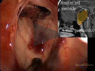
Watch an endoscopic craniopharyngioma tumor surgery
This video shows the endoscopic removal of a craniopharyngioma. The tumor is reached by working over the pituitary gland, taking extreme care to preserve the pituitary stalk. Once the tumor material is removed, the roof of the third ventricle (the deepest area where the tumor was) can be seen. The patient had intact pituitary function and vision after the surgery.
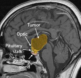
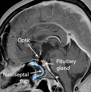
Large craniopharyngioma successfully removed using the Expanded Endonasal Approach.
You should ask your neurosurgeon if endoscopic surgery is appropriate for your tumor type and location, and always consider a second opinion. In some cases, an alternative minimally-invasive technique may be advantageous. UCLA neurosurgeons have extensive experience with all of the various techniques, including minimally-invasive "key-hole surgery" using the eyebrow to hide the incision.