Lung Cancer
What Happens After a Screen
Lung screening is performed with the goal of finding lung cancer in its early stages, before it causes symptoms. Lung CT's are reviewed by Radiologists who look for:
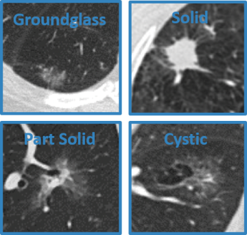
- Lung nodules: "spots" or "shadows" in the lungs. While lung cancer may appear as a nodule, the vast majority of nodules are NOT cancer. The Radiologist will describe the texture of the nodule, its size & recommendations for follow-up.
- Airway disease: signs of airways disease include abnormally enlarged or thickened airways as well as airways that are occluded/blocked.
- Emphysema: smoking can lead to destruction of lung tissue architecture.
- Fibrosis: lung scarring may be confined to one area or scattered throughout the lungs.
- Cardiac findings: while LDCT is not designed to evaluate the heart, calcification of the coronary arteries, aortic atherosclerosis and aortic aneurysms may be seen
- Findings outside of the chest: low dose CT of the chest is designed specifically to look at the lungs, however, abnormal findings in the chest wall or upper abdomen are sometimes seen. In this case, another imaging exam may be recommended to get a better look.
The Radiologist will write a report that summarizes important findings, and send it to your referring doctor. Each report includes recommendations for follow up. In most cases, this recommendation is for another screen in 12 months. In some cases, the recommendation may be to receive early follow up (1-6 months), a different type of imaging exam (e.g. CT Chest with contrast or PET/CT), or to consider a biopsy. In these cases, a multidisciplinary team will work to provide the care that you need.