Targeted, MRI-Guided Prostate Biopsy
Find your care
Trust our world-renowned prostate cancer experts to deliver your care.
For more information, connect with a prostate cancer care specialist at 310-794-7700.
Targeted Prostate Biopsy: What You Should Know
"Targeted biopsy" is being used at UCLA to diagnose prostate cancer. Targeted biopsy refers to using advanced imaging techniques to identify areas of the prostate suspicious for cancer, and then directly biopsying them. The traditional method of biopsy uses ultrasound imaging, with which tumors are hard to see. With targeted prostate biopsy, systematic sampling of prostate tissue is performed.
The new method employs sophisticated MRI (magnetic resonance imaging) technology, developed at UCLA, to visualize prostate cancer, and fusion of the MR images with real-time ultrasound using the specialized biopsy equipment. The result is a 10-20-minute procedure, done in the clinic under local anesthesia, that is much more accurate at finding significant prostate cancer in men.
A UCLA Team Approach to Targeted Prostate Biopsy
Targeted prostate biopsy was developed at UCLA by a multidisciplinary team, including members of the urology, radiology, pathology, and biomedical engineering departments.


The team was awarded a $1.7M grant from the National Cancer Institute to help bring the new fusion biopsy technology to full fruition. The first results of fusion biopsy at UCLA were published in 2013 (PDF). Team member Shyam Natarajan received a PhD degree in biomedical engineering, based largely on his work with the new method.
The targeted approach calls for an MRI to be performed before the biopsy of the prostate.
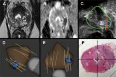
Using techniques of "diffusion-weighted imaging" and "dynamic contrast enhancement", in conjunction with a more powerful magnet (3 Tesla), many prostate cancers can be visualized on MRI images. By fusing these MRI images with real-time ultrasound, prostate biopsy can now be targeted directly toward specific areas of interest in the prostate. Published UCLA results show that targeted biopsy is several times more accurate than non-targeted, systematic biopsies at finding significant prostate cancer.
Value of Targeted Prostate Biopsy
This technology allows our team of specialists at UCLA to find hidden tumors missed by conventional prostate biopsy. Unlike all other major cancers, prostate cancer has proven difficult to image when still localized. This prostate-imaging technology offers a more exacting method to obtain biopsy specimens from suspicious areas within the organ. The targeted method has proven of great value for detection of tumors in men with prior negative biopsies, but persistently elevated PSA levels. It is also proving to be helpful in men enrolled in ASCAP, Active Surveillance of Cancer of Prostate (sometimes known as watchful waiting), providing the best possible information that a "low risk" tumor is just that.
Prostate Biopsy at UCLA: The Targeted Biopsy Process
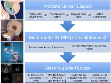
Patients who are suspected of having a prostate cancer diagnosis receive a multi-parametric MRI of the prostate after consultation with their UCLA urologist. An expert in prostate MRI will review the scan and provide an overall score to determine cancer risk. Using software created at UCLA, a 3-D image of the patient's prostate is then generated using the data and scores from the MRI prostate scans to clearly show the location of any suspicious areas.
The information is then ready for use in the clinic during a real-time ultrasound prostate biopsy at a later time. During the prostate biopsy, the MRI images are loaded into a3-D prostate biopsy system that allows the stored MRI images to be electronically transferred and fused with the real-time ultrasound, providing a 3-D image similar to a roadmap to help guide the tiny biopsy needle into targeted areas.
The work flow of MRI-guided TRUS biopsy, from patient selection to final prostate cancer diagnosis, as currently practiced at UCLA, is shown in this video.
Who Should Have a Targeted Prostate Biopsy?
A presentation of the initial Artemis experience at UCLA, establishing the biopsy-tracking accuracy of the device.
UCLA Urologists currently recommend it for men who are suspected of having prostate cancer, but who may have inconclusive tests results and have experienced any of the following:
- Persistent, unexplained elevated PSA, or
- Prior negative biopsy, or
- Increased prostate cancer gene expression (PCA3 urine test with an elevated score), or
- Apparent low-risk prostate cancer; interested in Active Surveillance (watchful waiting).
- All targeted biopsies are performed in the Urology, Westwood Adult clinic on an outpatient basis.
UCLA Advantages
UCLA's Prostate Cancer Program, affiliated with UCLA's Institute of Urologic Oncology and the UCLA Health Jonsson Comprehensive Cancer Center, is a multidisciplinary, comprehensive program offering the latest treatments for prostate cancer and compassionate care for its patients. Our program has a long-held team approach to treating prostate cancer that includes urologists, medical oncologists, radiation oncologists, pathologists, radiologists and clinical trial nurses that allows for specialized perspectives for deciding upon the best option for each individual patient.
Targeted Biopsy Marks Major Advance in Prostate Cancer Research
NBC News Story - www.nbclosangeles.com >