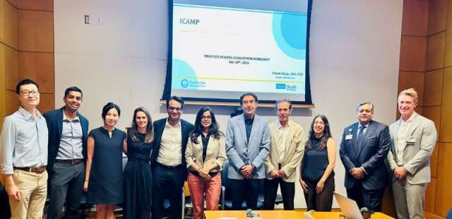
ICAMP: Imaging-Centric Abdominal Multiomics Program

Overview
The Imaging-Centric Abdominal Multiomics Program (ICAMP) Shared Resource generates, maintains and facilitates access to longitudinal, highly curated radiology-pathology data linked to clinical, molecular and outcomes information. Our purpose is to catalyze innovative research towards improving the imaging-based detection, diagnosis and treatment of cancer and advancing our understanding of the tumor microenvironment.
Our Vision
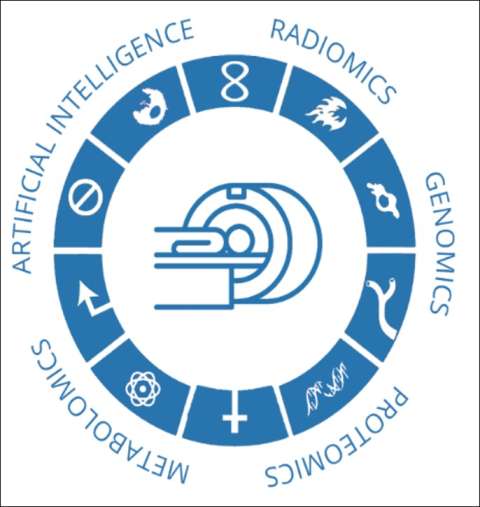
To develop an imaging-based multiomic model to understand and predict cancer biology and image-guided therapeutic outcomes.
Our Mission
Catalyze innovative research and tool development through data integration and curation to improve early detection, diagnosis and treatment of cancer.
- Acquire high-resolution, spatially registered radiology and pathology images.
- Capture high-value clinical and outcomes data.
- Spatially track and annotate findings across all observations at the lesion level.
- Collect biospecimens (blood, tissue) for multiomic analysis.
- Link with institutional resources such as the Institute of Precision Health, internal and external EMR and patient outcomes.
- Establish a web of multidisciplinary collaboration across radiology, pathology, genomics, and clinical care to accelerate precision medicine for abdominal cancers.
Explore
Regulation and Oversight
The Research Operations team oversees prospective consenting of participants, manages institutional review board protocols, and serves as an honest broker for data access.
Database and Integration
The Infrastructure team develops and maintains the database, data entry interface, dashboard, integration services and analytics services.
Image Archiving and Annotations
The Image Archiving & Annotations team is responsible for the banking, deidentification and annotation of radiological images for each domain.
Data Management
The Data Management team oversees data quality control activities and fulfillment of data access requests.
Type of Data Available
Clinical and demographical information
Radiological imaging (MRI, CT, ultrasound)
Digital pathology images
Image annotations (organ and lesion segmentation)
Expert-provided image interpretations (semantic features)
Measurements derived from images (radiomic features)
Outcomes information (disease-free survival)
Domains
Prostate
- Matched MRI and whole-mount resection cases
- Ex vivo imaging (a subset of resection cases)
- Prospective MR-guided biopsy cohort
Kidney
- Prospective biopsy cohort with prior multiphase contrast-enhanced CT and MRI scans
- CT or MRI cases with radiology pathology correlation
Liver
- US/CEUS/CT/MRI/quantitation of diffuse liver disease and liver neoplasms, with banked tissue and blood
ICAMP Prostate
ICAMP Prostate is a multidisciplinary cross-collaborative platform created to continuously improve MR/PET/Micro-US imaging with high-resolution histopathological and molecular correlations. Our vision is to unlock the potential of diagnostic imaging, enable detection of enable better patient triage and image-guided therapy, predict cancer biology, and allow for precise tissue acquisition and therapy.
Research Objectives
- MR, PET: Validate existing techniques and interventions, and develop and adopt new technologies with a focus on the correlations with pathology and microstructure
- AI: Deep learning, machine learning, and federated learning for prostate cancer diagnosis on imaging and digital pathology.
- Radiogenomics: Correlation with commercial tests and gene panels, correlation with general genomic panels, and differential gene expression based on PIRADS Score
Highlights and Key Publications
- New Techniques for Quantitative T2 and Diffusion MRI of Prostate Cancer
- Integrating MRI and Pathology to Characterize Prostate Tissue Microstructure and Improve Prostate Cancer Classification
- Personalized MRI analysis methods for accurate and precise DCE-MRI parameter estimation
- Artificial intelligence for improved diagnosis of prostate cancer
- Multimodal analysis of clinical and quantitative imaging data to improve prediction of biochemical recurrence
Recent Grants and Awards
- NIH/NCI R01 (PI: Sung/Wu) Integrating Quantitative MRI and Artificial Intelligence to Improve Prostate Cancer Classification
- UCLA Prostate Cancer SPORE (PI: Hsu) Computational Framework for Discovering and Validating Imaging Endotypes to Predict Clinically Significant Prostate Cancer Aggressiveness
- 2020 SAR Morton A. Bosniak Research Award
- 2020 SAR Trainee Scholarship Award
Overview
The Prostate ICAMP program has now migrated from a developmental phase to a mature phase where multiple databases have been built with clinical, pathological and MRI data with a mature interface, integration with the imaging biomarkers, and a mechanism for data and imaging retrieval. It is now a template for other programs including liver and kidney. We have finished the initial validation projects and are now implementing ongoing technical development as well as multiple artificial intelligence, federated learning and radiogenomic phases. We are looking forward to integrating the genomic and other biological data for expanded discovery in this new phase.
Available Prostate Data
| Cohort | Total Cases | Cases Annotated | Cases w/Clinical Data | Cases w/Tissue | Cases w/Blood |
|---|---|---|---|---|---|
| mpMRI Cases (incl Resection & Bxs) | 30,062 | 2,042 | 2,097 (all MRI data avail) | - | - |
| IPH Overlap | 6,695 | - | - | - | - |
| Resection Cases (MR & Path matches: micrographs available) | 1,649 | 1,604 | 1,649 | - | 95 |
| 3D Mold | 663 | - | - | - | - |
| Ex-Vivo | 111 | 110 | 49 | - | - |
| PSMA-PET (matched to Path, some MR) | 520 | 479 | 520 | - | - |
| microUS | 94 | - | - | - | - |
| HD WMHP Slides | 395 | - | - | - | - |
| Biobank MR-Guided Biopsies | 448 (20 re-consents) | 438 | 448 | 433 (18 w/tissue twice) | 126 |
ICAMP Kidney
The goal of Kidney ICAMP is to understand the imaging of small renal masses, explore the use of imaging for the differentiation of kidney masses, including benign and malignant tumors, and predict the biological behavior of renal cancers undergoing image-guided therapy.
Research Objectives
Our research objectives include reviewing the efficacy of the current imaging modalities available today in detecting and helping to characterize small renal masses and using AI to diagnose and report these lesions. We are evaluating outcomes in patients who were managed with surgery and ablative techniques like cryoablation, radiofrequency ablation and so on. We are also improving the prognostic determination of renal cell carcinoma (RCC) based on imaging features by building models or computer-based artificial intelligence diagnostic algorithms to enable CAD-based diagnosis of these lesions on the unique appearance of different types of tumors.
Highlights and Key Publications
- UCLA CT & MR Score for Classification of Solid Renal Masses
- Machine Learning-Based Quantitative CT Texture Analysis for Differentiation of Benign and Malignant Renal Masses
- Using Aorta-Lesion Attenuation to Differentiate Between Malignant and Benign Renal Lesions
- Validation of An Automated Software to Measure Total Kidney Volume in Patients with Autosomal Dominant Polycystic Kidney Disease
- Long-term outcomes for primary and metastatic renal cell cancers
Overview
Kidney ICAMP includes BioBank, Radpath and CT Perfusion projects, all of which are seeking to establish high-resolution, accurate spatial registration between radiology and pathology. The development of Kidney ICAMP has expedited over the past year. Researchers and staff have defined the scope of data collection and annotations, implemented new database initiatives and streamlined existing workflows.
Data Available
| Cohort | Total Cases | Cases Annotated | Cases w/Clinical Data | Cases w/Tissue | Cases w/Blood |
|---|---|---|---|---|---|
| Restrospective RCC Cases | 1,449 | 289 | 1,449 | - | - |
| Biobank CT/US Guided Biopsies (all consented) | 213 | 164 | 213 | 199 | 97 |
| CT Perfusions | 109 | - | 109 | - | - |
ICAMP Liver
The goal of Liver ICAMP is to integrate imaging, pathology, biospecimens, and molecular profiling to advance precision-medicine approaches for liver malignancies and metabolic liver diseases.
Research Objectives
Our research objectives include developing MR, MRI elastography, US, Contrast US, CT, and other image-derived biomarkers that predict tumor progression risk in hepatocellular carcinoma (HCC), correlating annotated imaging with clinical and pathologic data to support individualized risk assessment. We aim to evaluate histopathologic, genomic, and molecular correlates of tumor biology and treatment response following locoregional liver therapies, including thermal ablation, irreversible electroporation, and histotripsy. We also aim to develop non-invasive, blood-based biomarkers to assess progression risk in metabolic dysfunction-associated steatotic liver disease (MASLD) and its advancement to MASH and HCC, and build a validated patient-derived 3D liver organoid platform from cryopreserved biopsy tissue to enable individualized drug-response screening.
Highlights and Key Publications
- NIH/NCI U01CA230705 — UCLA Center for Early Detection of Liver Cancer (Zhou; Lu, co-I)
- NIH P30 Cancer Center Support Grant — Liver Disease Biobank collaboration (Han, Agopian)
- Multiple peer-reviewed publications, with additional abstracts, posters, and oral presentations at national meetings
Overview
Liver ICAMP integrates imaging, pathology, biospecimens, and molecular profiling in partnership with the Division of Hepatology & the Liver Cancer Center. The program collects imaging, biopsy-derived tissue, blood, and clinical data from patients with MASLD, MASH, hepatitis B/C, cirrhosis, and pre- and post-treatment liver malignancies, including hepatocellular carcinoma and cholangiocarcinoma. These specimens serve as a core resource for multimodal research to identify novel biomarkers and other factors associated with disease onset, progression, and outcomes, ultimately contributing to improved diagnostics, personalized therapies, and enhanced patient care.
Data Available
Banked Resources
| Category | Subtype | Tissue (biopsy) | Blood (initial) |
|---|---|---|---|
| Masses | HCC | 178 | 98 |
| Cholangio CA | 30 | 18 | |
| Others | 75 | 45 | |
| Diffuse liver disease | MASH | 30 | 20 |
| MASLD | 23 | 18 | |
| Others | 105 | 48 |
Faculty


Program Management & Oversight of Operations (PMO)


Data Management (QA)