Dear Doctors: I broke my ankle about 18 months ago. Now, in the area just above the break, the skin on my leg has turned a dark color, like a bruise. It doesn’t seem to be going away. Is there anything I can do to lighten this up? Do you think that laser therapy could help?
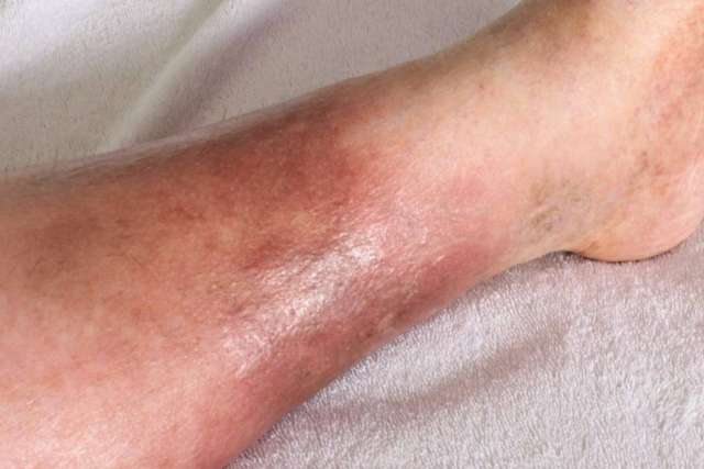
Dear Reader: You have described a condition known as “hemosiderin staining.” The term refers to areas of discolored skin that usually affect the lower leg, typically on the ankle and the top of the foot.
Hemosiderin staining occurs when the smallest blood vessels, known as capillaries, begin to leak. This can be due to a wound, a broken bone, a surgical incision or other types of trauma. It is also associated with certain illnesses that affect circulation. The most common of these is chronic venous insufficiency. This is a condition in which structural changes to the one-way valves in the veins prevent blood from circulating efficiently.
Both trauma and venous insufficiency result in the localized pooling of blood, which the body quickly begins to break down into its components. As the red blood cells are dismantled, the iron that they contain is released. This leads to the formation of a type of protein known as hemosiderin, which stores iron in the body. It’s the presence of iron that gives hemosiderin a rusty, brownish-yellow color. Over time, areas of hemosiderin staining can change color and intensify, becoming dark brown or even black.
In most cases, hemosiderin staining that has been caused by physical injury will gradually disappear as the damaged tissues heal. Depending on the individual, their general health and the nature and severity of their wound, it can take weeks, months or more than a year for the collected hemosiderin to disperse. In some cases, the pigmentation will become permanent.
Some people have success lightening the discolored skin with the use of prescription creams that contain hydroquinone. This is a compound that lightens the skin by interfering with the production of melanocytes, which are cells that produce pigment. While topical creams can prevent existing discoloration from becoming more pronounced, they may not completely remove it.

Laser therapy has also proven to be helpful at fading the discolored areas. For hemosiderin staining, a device known as a Q-switched laser is often used. This is the kind of laser used in tattoo removal. The Q-switched laser delivers short pulses of energy that are precisely calibrated to penetrate into the lower layers of skin. There, they destroy the pigment-containing cells. Because the sensation can range from uncomfortable to painful, a topical anesthetic is used. This type of treatment requires a series of visits to a dermatologist. It’s important to work with someone who specializes in this type of treatment.
In and of itself, the accumulation of hemosiderin is not a threat to health. However, because this can be a sign of a more serious underlying condition, it would be a good idea for you to be evaluated by your health care provider.
(Send your questions to [email protected], or write: Ask the Doctors, c/o UCLA Health Sciences Media Relations, 10960 Wilshire Blvd., Suite 1955, Los Angeles, CA, 90024. Owing to the volume of mail, personal replies cannot be provided.)