A new study led by UCLA Health researchers has identified a specific type of brain cell and a tiny structure on its surface that changes when the body is under stress, which may help explain why stress can lead to anxiety and depression.
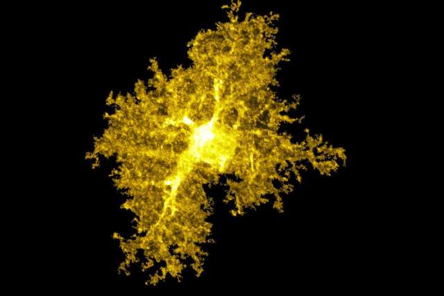
Published in the journal Nature, the research focused on astrocytes, bushy support cells in the brain, also known as glial cells, that have traditionally received far less attention than neurons in stress research. The team found that astrocytes in the amygdala, the brain region that governs fear and emotion, undergo measurable changes during stress. One notable change is the shortening of the cells’ primary cilium, an antenna-like protrusion that helps cells sense and respond to their surroundings.
“We've mostly thought about stress and mood disorders in terms of neurons firing signals to each other,” said the study’s co-first authors Sara Gutierrez Pelaz, a postdoctoral researcher for UCLA professor of physiology and neurobiology Baljit Khakh at the David Geffen School of Medicine at UCLA and Katsukuni Mitsui who was a visiting scientist in Khakh’s lab from ONO Pharmaceutical Co., Ltd. “Our findings show that a different type of brain cell, and a structure on that cell most people have never heard of, also plays a role in how the brain responds to stress.”
Using mouse models of chronic stress, the researchers observed that amygdala astrocytes showed altered gene and protein levels, some of it tied to primary cilium function, along with physically shorter cilia. Researchers restored cilia through two different approaches: a lab technique that switches specific receptors on astrocytes on or off, and a drug that targets a receptor called S1PR1. The study found these treatments reversed many of the molecular changes and improved some stress-related behaviors in the mice, including signs of anxiety and reduced interest in normally pleasurable activities.
Notably, the S1PR1 receptor is also found in human amygdala tissue, and it is already the target of an FDA-approved multiple sclerosis drug called ponesimod. When the researchers gave that drug to stressed mice, it produced similar improvements, suggesting a potential route to testing the approach in people.
“Because the receptor involved is already targeted by an approved drug, this gives us a realistic starting point for exploring new potential treatment approaches, rather than starting from scratch,” said Baljit Khakh, the study’s senior author.
Researchers also examined gene activity in brain tissue from people with major depressive disorder, bipolar disorder and psychosis-related disorders. They found that cilium-related genes in astrocytes were disrupted in those conditions as well, reinforcing the findings in the mice study.
Stress-related conditions such as anxiety and depression affect a large share of the population, and current treatments do not work for everyone. By identifying a specific cellular structure that changes with stress and can be restored, the study offers a new angle for developing treatments, one focused on glial cells rather than neurons alone.
The findings are early-stage and based on animal models. More research will be needed to determine whether targeting astrocyte primary cilia could become a viable treatment strategy in humans.