For patients with debilitating spine conditions that fail to improve with nonsurgical treatments, the UCLA Spine Center offers an endoscopic procedure that is less invasive than both traditional open surgery and minimally invasive approaches using a microscope. The result, says Don Y. Park, MD, is better visualization of the spinal anatomy during surgery, along with faster recovery and a lower risk of complications.
Dual portal endoscopic spine surgery is a potential option for patients with the most common conditions Dr. Park sees, including herniated disks — the slippage, rupture or bulging of the rubbery cushions along the spine, often affecting young adults; lumbar spinal stenosis, a narrowing of the spinal canal, typically from age-related wear; and spinal spondylolisthesis, a slippage in one of the vertebrae that causes instability. Dr. Park notes that the new approach, which was developed in South Korea and where he trained to perform the procedure, is driven by a demand among patients and surgeons for less-invasive spine procedures. “This is the next evolution of minimally invasive spine surgery,” he says.
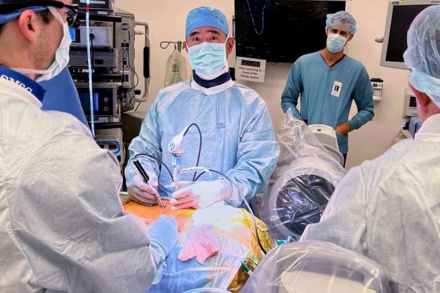
With traditional spinal operations, surgeons can directly visualize the anatomy, but the larger incisions increase the risk of infection and blood loss while increasing pain and lengthening recovery time. The open approach has increasingly given way to minimally invasive spine surgery, involving incisions of 18-20 millimeters and the use of tubes and microscopes to visualize the spinal anatomy. With the transition to endoscopy, the surgery is performed through even smaller incisions — less than one centimeter — and cameras are employed instead of microscopes to provide better visualization.
“The complications of surgery are often due to inadequate visualization, which has been one of the critiques of minimally invasive surgery,” Dr. Park says. “With dual portal endoscopic spine surgery, we can zoom in with the camera, just as you would with arthroscopy,” in which cameras are placed in the joints of the knee, shoulder or hip. “We use water rather than a tube, which allows the surgeon to drive the camera closer to the anatomy and see a level of detail that is impossible with a microscope.”
As with arthroscopic techniques, dual portal endoscopy utilizes two incisions — one of approximately 3-to-4 millimeters for the camera, and a second 6-to-7 mm incision for the surgical tools. Decoupling the camera and the surgical equipment provides a more expanded view along with greater flexibility in positioning the instruments, Dr. Park explains.
In six-week follow-ups with individuals who have had dual portal endoscopic spine surgery at UCLA, patients have shown significant improvements in their pain and disability measurements — results similar to those reported in South Korea, which has a longer track record with the approach.
UCL A is currently the only academic center in California offering this type of surgery. “People with these conditions are often in substantial pain and unable to walk for long distances or perform the activities of daily living,” Dr. Park says. “Many studies have shown that those who are having continued symptoms despite nonsurgical treatments can experience significant improvements after surgery. It’s exciting to be able to offer a procedure that provides those results while allowing them to bounce back much more quickly.”