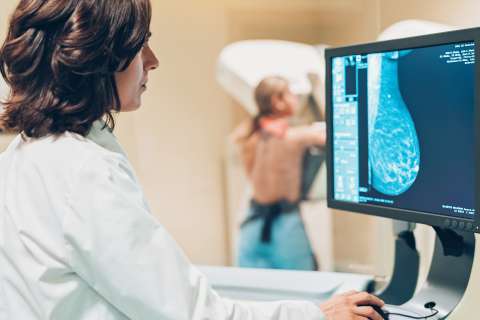
Breast Imaging
Our team leads the way in advanced mammography and breast imaging in Los Angeles. We offer a wide range of state-of-the-art screening and diagnostic tools.

Why choose UCLA Health for breast imaging?
Since we opened the first mammography center in Los Angeles, UCLA Health has been at the forefront of women’s health and breast imaging services. We were the first center to become accredited by the Mammography Accreditation Program and, more recently, the first facility in California to become 100% digital.
Highlights of our program include:
Multispecialty, team-based treatment: We offer whole-person care from a team of experts. Our team includes breast radiologists, breast surgeons, radiation oncologists, pathologists, genetic specialists, nurse practitioners and registered nurses. We tailor screening, prevention and risk-reduction strategies to your needs to deliver the best possible outcome.
Easy access to care: We offer women’s imaging and breast imaging services in seven locations, so you have easy access to care close to home. Each of our facilities provides a broad spectrum of state-of-the-art tools, ensuring the highest level of quality for screening, diagnosis and treatment.
History of innovation: Our specialists are recognized leaders in breast imaging. Not only were we the first center in Los Angeles to offer breast ultrasound, but we were also among the first in the region offering 3D mammography technology. Today, we are a totally digital mammography facility, providing high-quality care in a relaxing, compassionate environment.
Breast conditions we treat
The primary goal of breast imaging is to detect breast cancer in its early stages, when it’s easier to treat. Breast imaging can also detect noncancerous (benign) conditions, such as fluid-filled growths (cysts) or fibrocystic breasts.
Screening mammography is one of the most important tools for breast cancer detection. Since screening mammograms became widespread in the mid-1980s, the national breast cancer mortality rate has declined by 40%. The American College of Radiology recommends a few guidelines for screening mammograms:
Women over 40 with an average risk of breast cancer should have a screening mammogram each year.
Women under 40 with a high risk of breast cancer may start annual screening mammograms, and possibly yearly MRIs, at their doctor’s guidance. Factors that increase your risk of breast cancer include:
- Family history of breast cancer
- Having dense breasts or a personal history of breast cancer
- History of chest radiation at a young age
- Mutations in the BRCA1 or BRCA2 genes
- Starting menstrual periods younger than 12 or starting menopause after 55
Tests and procedures we offer
Our specialists offer a wide range of tests and tools to screen for and accurately diagnose breast cancer.
Breast imaging exams
Types of breast imaging we offer include:
3D mammograms (breast tomosynthesis): Mammograms are low-dose breast X-rays. A technician takes X-rays from several views and a computer compiles those images into a 3D picture that allows us to see breast tissue clearly.
Screening mammogram: We perform screening mammograms for patients who have no signs of breast cancer. These tests involve two views of the breast. Screening mammograms are one of the best tools we have for finding cancer in early stages.
Diagnostic mammogram: If a patient has symptoms that could point to breast cancer, we do a diagnostic mammogram. It involves several views of the breast and focuses on particular areas of concern. We can use these tests to diagnose the cause of breast changes, such as an unusual lump, pain or nipple discharge.
Breast ultrasound: Ultrasound uses high-frequency sound waves to generate breast tissue images. We may use ultrasound in addition to a mammogram to get a closer look at breast changes. For example, an ultrasound can help us differentiate between a fluid-filled cyst, which is highly unlikely to be cancer, and a solid mass, which may need further testing. Ultrasounds are also often useful for women with dense breast tissue, for whom mammograms may not be as reliable.
Breast MRI: We often use MRIs for women who have been diagnosed with breast cancer. These tests can help measure the size of a mass, look for other tumors or check for tumors in the opposite breast. MRIs use magnets and radio waves to produce images of the breasts in all directions: top to bottom, left to right and front to back. Some women with a high risk for breast cancer may benefit from screening MRIs along with yearly mammograms.
Breast biopsies
When breast imaging detects a lump or other irregularity in breast tissue, it doesn’t necessarily mean you have breast cancer. We perform a biopsy to check breast tissue for cancer.
We also use several techniques to evaluate breast tissue, including:
Surgical biopsy: You receive local anesthesia to numb your breast along with intravenous (IV) sedation to help you relax. Then, a surgeon makes incisions to remove a small portion of breast tissue or the entire tumor. They usually close the incision with stitches and place a sterile dressing over the incision site.
MRI- or ultrasound-guided breast biopsy: Your provider places a hollow biopsy needle into the breast, then uses MRI or ultrasound images to confirm that they have the needle in the correct location. They obtain the tissue sample through the biopsy needle, bypassing the need to make an incision in the breast.
Stereotactic breast biopsy: Providers use mammography to pinpoint the location of the lump or breast change. Then, they use a biopsy needle to obtain a small tissue sample.
Presurgical wire localization: Using ultrasound or mammography guidance, a radiologist places a small wire in the exact location of the abnormal tissue. This wire serves as a “roadmap” for the surgeon to find and remove cancer. It is inserted the day of surgery and removed at the end of the procedure.
Meet our team
Our breast imaging team uses state-of-the-art, research-driven tools to screen for and diagnose breast cancer. We build on a long history of innovation to bring patients the most advanced technology to detect breast cancer.
Contact us
Call 310-301-6800 to request an appointment with a breast imaging specialist at UCLA Health.
Find your care
We offer the full breadth of services to screen for and diagnose breast cancer. To learn more about breast imaging services, call 310-301-6800.