PSMA PET/CT Imaging
Why choose PSMA PET/CT Scan for Prostate Cancer?
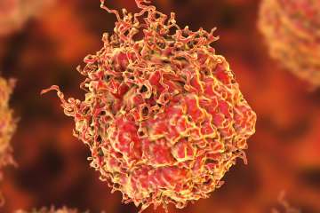
Precisely locating prostate cancer so that it can be treated is extremely challenging. This is because prostate cancer tumors can be found not only in the prostate but also throughout the pelvis and the body in cases where the tumors have migrated.
Effectively pinpointing and eliminating these tumors is important because prostate cancer is the second most common cancer in men and the second leading cause of cancer death in men.
Find Your Care
You can trust UCLA Health to deliver an accurate diagnosis and outstanding care using the latest advances in nuclear medicine. Call 310-794-1005 to connect with an expert.
In 2022 alone, an estimated 268,490 cases of prostate cancer will be diagnosed, with about 34,500 deaths
For this reason, UCLA Health and UC San Francisco validated for use in the United States a diagnostic innovation known as the prostate-specific membrane antigen or PSMA PET/CT scan. This scan is approved by the U.S. Food and Drug Administration and it enables more precise detection of prostate cancer for treatment planning and targeted care.
PSMA PET/CT is a better standard of care because it is more accurate
UCLA Health researchers, along with colleagues at UC San Francisco, studied PSMA PET/CT for several years. These studies led to the FDA approval of this imaging technique’s use in the United States.
PSMA PET/CT scans for prostate cancer provide better care for men who are initially diagnosed with prostate cancer or for men who were previously treated but have experienced a recurrence of the cancer. In both cases, PSMA PET/CT is better able to find the extent of this cancer in the body.
PSMA PET/CT enables more precise detection of prostate cancer
PSMA PET/CT works using a radioactive tracer, called 68Ga-PSMA-11, which is manufactured on site at UCLA Health. Nuclear medicine physicians inject this tracer which attaches itself to proteins known as prostate-specific membrane antigens (PSMA). Because prostate cancer tumors overexpress these proteins on their surface, physicians can pinpoint their location with the tracer.
The current standard of care in prostate cancer imaging is a technique called fluciclovine PET/CT, which involves physicians injecting patients with a synthetic radioactive amino acid.
In their research comparing PSMA and fluciclovine PET/CT, the UCLA and UCSF research teams found that imaging with PSMA PET/CT was able to detect significantly more prostate lesions than fluciclovine PET/CT in men who had undergone a radical prostatectomy but had experienced a recurrence of their cancer.

PSMA PET/CT was validated for use in the United States by UCLA and UCSF. The UCLA research team was led by the nuclear medicine faculty of the Ahmanson Translational Theranostics Division of the Department of Molecular and Medical Pharmacology. They worked in collaboration with the Departments of Urology, Radiation Oncology and Radiology, along with support from the David Geffen School of Medicine at UCLA, the UCLA Health Jonsson Comprehensive Cancer Center and the Prostate Cancer Foundation.
How to receive a PSMA PET/CT scan and prostate cancer care at UCLA Health
UCLA Health provides comprehensive and customized care for men with prostate cancer. Our physicians use the PSMA PET/CT scan alongside radiation therapy, chemotherapy, surgery, and all other treatment modalities offered to make sure men receive the best possible care – from diagnosis to treatment to follow-up.
To ensure the best treatment possible, UCLA Health’s nuclear medicine physicians, medical oncologists, radiation oncologists, radiologists, urologists and surgeons optimize care for each person receiving treatment. The prostate cancer care at UCLA Health is backed by multidisciplinary tumor boards with physicians from different specialties and subspecialties, along with genetic counselors and representatives from allied health services. Together, these specialists and experts meet once a week to talk about each new cancer patient and the path forward for their specialized treatment.
How to get started with a PSMA PET/CT Scan for prostate cancer
Referrals
UCLA Health welcomes patient referrals for a PSMA PET/CT scan from physicians who are based anywhere in the world. For patients who have questions about gaining a physician’s referral, please contact us.
Connect with us
For an appointment, call us at 310-794-1005. Appointments are taken Monday through Friday, 8 am to 5 pm. Please leave a message after hours.
Internal healthcare providers can e-mail the Nuclear Medicine Clinic at UCLA Health or place an order in CareConnect for individuals who are already patients.
Where treatment happens
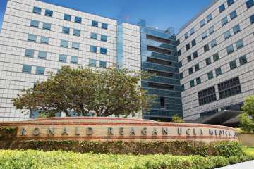
UCLA Health’s PSMA PET/CT scan is performed at the Ronald Reagan UCLA Medical Center on UCLA’s campus in the Westwood area of Los Angeles.
Once the PSMA PET/CT scan is complete, men can receive prostate cancer care at UCLA Health’s hospitals in Westwood and Santa Monica, or at UCLA Health’s Encino medical office. The scan can also be used in conjunction with care at any other hospital where the patient is being treated.
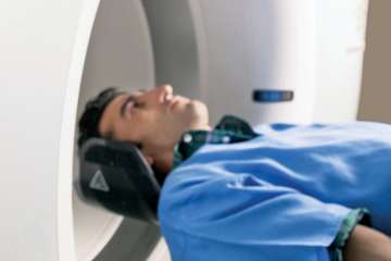
The total time it takes for a PSMA PET/CT scan is about two hours. What will happen is the tracer will be injected by a UCLA nuclear medicine technologist. Then the patient waits one hour before entering the PET/CT scanner for 30 minutes.
Dr. Johannes Czernin and Dr. Jeremie Calais with UCLA’s nuclear medicine team helped lead the clinical trials for the use of PSMA PET scan imaging in the United States. Team members also include Drs. Robert Reiter, Matthew Rettig, Nicholas Nickols and Amar Kishan.