Gout Care
Our specialized rheumatologists provide targeted, research-based gout care. We participate in studies and clinical trials to bring you the latest effective diagnostic tools and treatments.

Why choose UCLA Health for gout care?
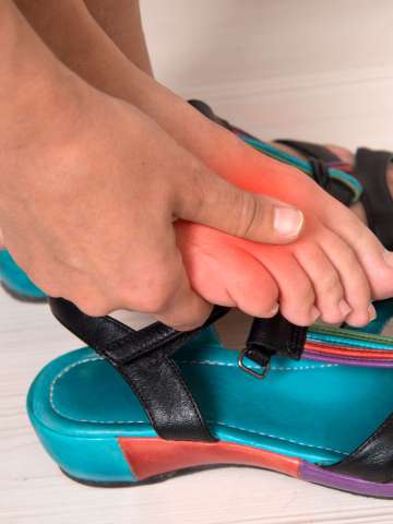
Gout is the most common type of inflammatory arthritis. It occurs due to a buildup of uric acid and causes bouts of pain, often affecting your big toe. At UCLA Health, our rheumatologists provide comprehensive gout treatment to help you find relief.
Highlights of our program include:
Research focus: Our gout specialists are active researchers who study innovative ways to diagnose and treat gout. One of our rheumatologists was the principal investigator for the American College of Rheumatology Gout Guidelines and Quality Improvement Measures. We have also developed a new lens-free microscope that improves visualization and efficiency when diagnosing gout.
Specialized expertise: UCLA Health rheumatologists have specific expertise in gout treatment, leading to specialized, advanced care. Our program includes physicians certified in musculoskeletal ultrasound technology by the American College of Rheumatology. These in-office musculoskeletal ultrasounds are crucial for both identifying signs of gout and treating it by removing fluid from joints.
Teaching and training: As an academic health system, we focus on training the next generation of physician-leaders. Our rheumatologists teach best practices in gout treatment to medical students, residents and fellows.
What is gout?
Gout is a type of inflammatory arthritis that affects more than 8 million adults in the United States. It occurs when levels of uric acid, a waste product in your blood, build up. As a result, urate crystals form and deposit in your joints. Gout usually affects one joint at a time.
Urate crystals can cause attacks of inflammation and severe pain, most frequently in the big toe. Gout is more common in men, but it also affects women after menopause.
Uric acid builds up when your body doesn’t eliminate enough of the waste product through your kidneys. Usually, this is genetic. However, your diet, body size and some medications can affect your gout risk. Many people with gout also have high blood pressure, high cholesterol or diabetes.
Gout tests and treatments we offer
UCLA Health rheumatologists diagnose gout using sophisticated tools. With an accurate diagnosis, we can create a targeted treatment plan for you.
Gout diagnosis
To diagnose gout, we typically use tests such as:
Blood tests: Your provider may use a blood test to check your uric acid levels. Typically, people have uric acid levels between 3.5 and 7.2 milligrams per deciliter (mg/dl). If your uric acid levels are above 6 mg/dl, your provider may order additional tests to check for gout.
Imaging tests: Your provider may use ultrasound or a specialized CT scan called a dual-energy CT scan to look for gout. An ultrasound uses high-frequency sound waves to identify changes in your soft tissues that could indicate gout. A dual-energy CT scan uses two X-rays of varying intensity to create detailed images. It can help your provider analyze urate crystal deposits more closely. UCLA Health researchers may also use dual-energy CT scans to analyze gout in comparison to your cholesterol levels. Evaluating gout and cholesterol can help reduce the risk of heart attack or stroke, which is typically higher for people who have gout.
Fluid analysis: Your provider takes a small sample of fluid from your affected joint and analyzes it under a microscope to look for urate crystals. Our researchers have created a crystal lens-free microscope, which allows us to perform the same analysis in less time with a larger field of view and higher resolution.
Gout treatment
Most people with gout need to take medications to control gout. Some medicines, such as nonsteroidal anti-inflammatory drugs (NSAIDs) or steroids may provide short-term relief from gout attacks. Most people need a daily, long-term medication to reduce urate crystals and prevent future attacks. Your rheumatologist can recommend the best medication for your needs.
Meet our team
UCLA Health rheumatologists provide targeted, comprehensive treatment for gout. We are active researchers who bring you the latest diagnostic tools and effective treatment options.

Contact us
Call 310-825-2448 to request an appointment with a gout specialist in Santa Monica or 310-582-6350 to request an appointment in Westwood.
Find your care
We have specific expertise and training in gout diagnosis and treatment. To learn more about gout care at UCLA Health, call 310-825-2448 for our Santa Monica office or 310-582-6350 for our Westwood office.
Support Our Work
If you would like to learn more about ways to give, please contact our Senior Executive Director of Development, at 310-794-4746. Thank you very much for your interest!