Breathing Motion Modeling in Radiation Therapy
5DCT Respiratory Motion Modeling
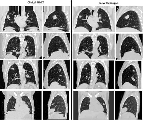
Accurate time-resolved imaging is crucial to the management of respiratory motion in radiation therapy treatment planning. Conventional techniques for producing breathing gated images commonly suffer from artifacts due to their inability to account for irregular breathing cycles. A novel CT acquisition and analysis technique, 5DCT, is utilized at UCLA in order to better inform treatment plan design for mobile tumors. Compared to current commercial protocols, 5DCT offers several advantages:
- Images do not contain artifacts that arise from the sorting of images performed by commercial techniques.
- Any number of images at user-selected breathing phases can be generated without altering the acquisition protocol.
- Resulting images are low-noise while patient dose remains similar to or less than current techniques, and the entirety of the imaging dose potentially contributes to image quality.
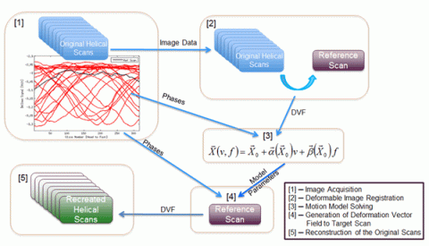
5DCT exploits standard fast helical acquisition with simultaneous breathing surrogate measurement, deformable image registration, and a breathing motion model to remove sorting artifacts. The accuracy of the motion model is assessed by reconstructing the original fast helical scans, which provide a ground truth.