Ureteropelvic Junction (UPJ) Obstruction
Find your care
We deliver customized urology care based on your unique needs. To learn more or connect with a urology specialist, call 310-794-7700.
What Is UPJ Obstruction?
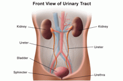
Ureteropelvic junction (UPJ) obstruction is a partial or total blockage at the place where the organ that produces urine (the kidney) and the tube that carries it to the bladder (the ureter) are joined. The kidney filters blood and collects urine at its center, where the renal pelvis funnels it into the ureter so that it can be delivered to the bladder. More common in children than in adults and often resulting from a congenital abnormality, UPJ obstruction impedes the flow of urine, causing it to build up and resulting in hydronephrosis – an enlargement of the renal pelvis – and the potential for kidney damage.

UPJ obstruction is the most common cause of hydronephrosis detected on prenatal ultrasound or in newborns. The blockage can range from minimal to severe. Mild cases usually don’t damage the kidney or impair its function, but they can predispose the child to urinary tract infections. Severe cases can cause significant harm to the kidney.
UPJ Obstruction Causes, Symptoms & Diagnosis
UPJ obstruction is often diagnosed during prenatal ultrasound, when the enlarged kidney is seen. For those that occur later or are not detected at birth, symptoms suggesting UPJ obstruction include hematuria (blood in the urine), urinary tract infection, kidney infection, kidney stones, and abdominal discomfort.
While the most common type results from a narrowing of the ureter as it forms in fetal development (usually because of an abnormality in the development of the muscle surrounding the UPJ), UPJ obstruction can also occur later in life and can be caused by other factors, including compression of the ureter by inflammation, kidney stones, scar tissue, abnormal blood vessels, or a tumor. Diagnostic tests help to determine the degree of UPJ obstruction and whether surgery is necessary.
Treatment for Ureteropelvic Junction Obstruction
When the obstruction is mild, it is usually left to correct itself. Antibiotics may be used to prevent infection, and the child is monitored every 3–6 months with a renal ultrasound. Because of the potential for kidney damage, more severe cases tend to require pyeloplasty, a surgical procedure that removes the blockage and reconnects the ureter and the renal pelvis. The success rate is higher than 95 percent, and the procedure can often be done laparoscopically. The child continues to be followed even after successful repair to ensure proper kidney function.