When a patient comes into a trauma center at a large U.S. hospital with a potentially life-threatening injury, doctors can begin looking inside their body in a matter of minutes.
In a typical trauma bay in Southern California, a large ultrasound machine can be rolled to the patient’s bedside almost immediately. Right away, it can detect hidden internal bleeding or other life-threatening conditions that may not be apparent during a physical examination.
But, that level of rapid diagnostic care is not available in many parts of the world and even some parts of the United States, where roughly 30% of the population lives in what are known as “trauma deserts” – communities where patients don’t have access to high-quality trauma care. Around the world, the problem is far greater.
Ariane Christie, MD, a trauma surgeon and researcher at the David Geffen School of Medicine at UCLA, along with colleagues stateside and in West Africa, has been exploring some of these challenges for the last decade.
And these were the questions she and her fellow investigators chose to explore: What if the technology used to find these hidden injuries could be made small enough to fit in a fanny pack? And what if the training required to use that technology could be reduced to just a few hours?
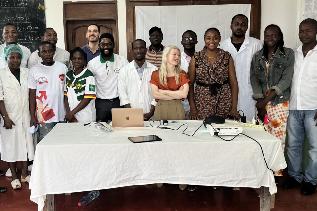
These questions also formed the basis of an international research program funded by the National Institutes of Health (NIH) to bring portable ultrasound to places where conventional imaging can be challenging or even impossible to access. Specifically, Dr. Christie, along with UCLA trauma surgeon Catherine Juillard, MD, and others, have been working to bring portable ultrasound imaging to hospitals in Cameroon, Ghana and Kenya, in addition to training doctors and medical students there about how to use this technology.
Turning a large machine into a portable tool
Dr. Christie says over the last 20 years, ultrasound technology has undergone a dramatic transformation with several companies developing small, ultrasound probes that connect to smartphones.
“The probes are the parts that touch the patient and the other side of that plugs into the phone,” she said. “It provides an image at a fraction of the cost of a large ultrasound machine, which makes them much more affordable. They are also far more portable, which means they can be transported from country to country or from one area of the hospital to another with much greater ease.”
But getting the device and the technology to these places was only part of the problem. The other piece of the puzzle was getting medical professionals trained to use them. Cameroon, where much of Dr. Christie’s project is based, illustrates this daunting challenge.
“In Cameroon, the only people who know how to read an ultrasound are radiologists, and there is one radiologist for every 500,000 people in that country,” she said. “There simply aren’t enough experts to stand in emergency departments and teach every physician how to perform and interpret ultrasound examinations.”
Teaching a complicated skill in a few hours
Dr. Christie’s passion for global health and injury care goes back over a decade. She began working in sub-Saharan Africa as early as 2012 and later spent a year living in Cameroon, from 2016 to 2017. It was when she began her trauma fellowship at the University of Pittsburgh in 2020 that she realized portable ultrasound could be paired with a radically shortened training program.
So, instead of trying to turn doctors into ultrasound specialists, the goal would be to teach them a specific set of examinations to help identify life-threatening conditions. The research team at UCLA Health, along with partners in Cameroon, spent the first year developing a curriculum to do exactly that. The training would not only teach physicians how to get the specific ultrasound views needed to identify critical injuries but also how to interpret what they see.
“It’s not useful if you’re able to get the views, but you don’t know what you’re looking at,” Dr. Christie said.
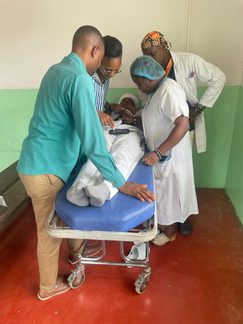
The curriculum they developed essentially took a provider with no previous ultrasound experience from the beginning to competency in about five hours, she said.
“Another challenge is because there are so few providers we can’t actually take them away from their environments for very long because people need healthcare,” Dr. Christie said. “So, we knew that we needed to be able to teach this very quickly.”
How these ultrasound exams can save lives
It only takes a minute or two for a physician to scan an injured patient’s body with these portable devices. Dr. Christie says one of its most important functions is detecting internal bleeding.
“A patient can have a significant amount of blood collecting in their belly or chest, which might not be obvious in a physical exam,” she said.
But, a portable ultrasound can provide immediate indication that something is wrong. This technology can also detect a pneumothorax, commonly known as a collapsed lung, Dr. Christie said. When this happens air escapes from the lung and accumulates in the surrounding space, compressing the lung. In severe cases, this could interfere with heart function and circulation.
“In these cases, we could simply make a small opening in the chest to release the trapped air and save the person’s life,” she said. “But, the clinician has to first recognize the problem.”
Another potentially fatal condition that can be detected with ultrasound is cardiac tamponade. This is when an injury causes blood to collect in the pericardium – the sac surrounding the heart. This pressure can prevent the heart from beating normally.
“Again, this is a totally treatable condition,” Dr. Christie said.
How the investigations have evolved
The first clinical trial offered an important proof of concept. Over a six-month period across three hospitals in Cameroon, the research team dramatically increased the percentage of patients receiving ultrasound imaging from 6% to 80%.
The question they are looking into now is whether the program actually changed outcomes for patients. Investigators are looking into how accurately physicians interpret the ultrasound exams in real-world clinical settings and whether the technology changes the diagnosis and treatment of critical injuries.
Dr. Christie’s interest in Cameroon grew out of a broader commitment to global health. While she had begun working in sub-Saharan Africa more than a decade ago, her subsequent work in Francophone Africa drew attention to particularly poor injury outcomes.
Cameroon became a natural partner because the country’s traumatic injury death rate is about 113 deaths per 100,000 people – more than twice the U.S. rate of about 42 per 100,000. Dr. Christie and Dr. Juillard had already established longstanding relationships in Cameroon and in 2022, they wrote the grant for the ultrasound project with their Cameroonian partner, trauma surgeon Alain Chichom Mefire, MD.
The first development phase took place in 2023 and 2024. The six-month clinical trial followed in 2024-25. The researchers met their initial clinical metrics in the summer of 2025, qualifying the project for an additional three years of NIH funding. The newest implementation phase is scheduled to run from the winter of 2026 through the summer of 2028, with the initial implementation taking place in Cameroon.
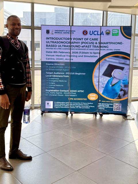
The project is expanding to Ghana and Kenya as well, Dr. Christie said. In Kenya, preliminary results show providers may be able to reach competency using the curriculum in about two hours. The program has also attracted interest from the West African College of Surgeons. In early 2026, the team traveled to Ghana to test the curriculum with 26 Ghanian physicians; 25 completed the competency testing successfully the same day, Dr. Christie said.
“Ultrasound can be used for many other medical purposes including evaluating pneumonia, heart examinations, helping clinicians obtain vascular access and assessing obstetric conditions,” she said, adding that this essentially means portable ultrasound could become something more than a trauma intervention. It could become a general diagnostic tool for communities where access to advanced imaging remains limited.
Speedier diagnostics can promote health equity
Dr. Christie’s team is considering testing the program at UCLA Health to determine whether the curriculum could benefit surgical trainees here.
“Residents already have an enormous amount to learn,” she said. “If a skill that traditionally takes months could be taught within a matter of hours, that could be extremely valuable.”
These portable ultrasound devices could be life-saving in U.S. trauma deserts, too, she said, by providing additional diagnostic capability where advanced imaging may be hours away.
Most people go into the healthcare research field driven by a love of investigation and to genuinely help people, Dr. Christie said.
“Anyone who studies public health for any length of time comes to the realization it’s critical to deliver high-quality care to the individual by making changes in the system,” she said. “If we’re successful with this project we’ll hopefully have the opportunity to be involved in the improvement of healthcare for many, many more people than I’ll ever treat.”
Promoting health equity is a big part of it, she said.
“It’s just random where you’re born,” Dr. Christie said. “So much of your options and how you’ll do if something bad happens to you is just a factor of where in the world you are born and what the system is there.”
She believes there is no reason such gross inequities should exist in this era of rapid technological advancements.
“We need to make sure that those opportunities are delivered more universally to improve outcomes for everybody,” Dr. Christie said. “It really is better for the entire world.”