The National Cancer Institute (NCI) has renewed, for a second five-year period, a $10-million grant for an innovative molecular imaging center at UCLA, officials announced today. The new funding, bringing UCLA's total support to $20 million, will allow the scientific advances made in the last five years to be translated into the clinic in the next five years to improve the diagnosis and staging of cancer.
The grant funds the UCLA Center for In Vivo Imaging in Cancer Biology, one of the NCI's specialized In Vivo Cellular and Molecular Imaging Centers (ICMICs). The first such facility on the West Coast, UCLA's imaging center brings together the expertise and experience of researchers from UCLA's Molecular and Medical Pharmacology department, the Crump Institute for Molecular Imaging and UCLA's Jonsson Cancer Center.
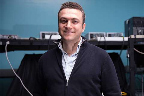
Harvey Herschman, principal investigator of the UCLA imaging center, said the goal of the initial $10-million, five-year grant was to extend the development of non-invasive molecular imaging technologies pioneered at UCLA, and make these new technologies part of the regular "toolbox" used by molecular and cell biologists at UCLA who are studying cancer in animal models.
"We accomplished these goals. Dozens of research labs on campus now use these technologies," said Herschman, director of basic research at UCLA's Jonsson Cancer Center and a professor of biochemistry and pharmacology. "For the next five years, our objective will be to translate into the clinic these new technologies and discoveries to improve diagnosis and staging of cancer, and to much more rapidly distinguish those patients who might respond to certain therapies from those who would not. If we can do that, patients can receive the appropriate therapy for their cancer as soon as possible."
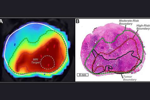
One project focuses on optimizing positron emission tomography (PET) imaging technologies to rapidly monitor metabolic responses of brain and lung tumors to experimental therapies. Researchers hope to identify those tumors sensitive to a drug by showing a metabolic response and distinguishing them from non-responders. Metabolic measures of response are detectable much earlier than the structural changes in tumors that are usually measured in current clinical procedures, so doctors can tell more quickly if a drug is working.
Another translational project involves the use of reporter genes to mark modified immune cells that will be used to stimulate the body's own response to melanomas. Researchers will be able to trace the reporter genes, using imaging, and determine the location of the injected immune cells, their ability to target tumor tissue, their longevity in patients and their biological activity.
UCLA received one of the first ICMIC awards in 2001, along with Harvard University and Memorial Sloan-Kettering in New York. Currently, there are seven such molecular imaging centers in operation across the country. UCLA is the first institution to be awarded a renewal of an ICMIC grant, Herschman said, and to receive a second $10 million funding commitment.
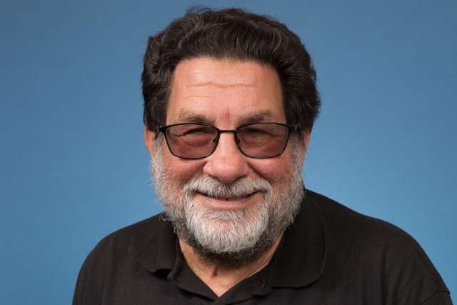
Judith C. Glasson, director of the Jonsson Cancer Center and a professor of medicine and biological chemistry, said discoveries made in UCLA's molecular imaging center hold tremendous potential for cancer research and patient care.
"These discoveries will hasten development of safe, effective treatments for patients by allowing researchers to more rapidly and thoroughly evaluate the benefits and limitations of certain experimental therapies," Gasson said.
Research at the UCLA molecular imaging center already has shed new light on the safety and effectiveness of gene therapy and other gene-based treatments. Investigators also are studying how cancer develops, grows, spreads, forms its own blood supply for nourishment and interacts with the human immune system.
Herschman said understanding cancer at its molecular and cellular levels plays a key role in improving diagnosis, treatment and prevention.
"Being able to non-invasively see what is going on in the body will give us the information necessary to overcome hurdles such as not being able to predict a tumor's behavior or not knowing how a tumor will respond to certain treatments," he said.
NCI Renews $10 Million Grant for Molecular Imaging Center for Cancer Research
Related Content
Share: