Story highlights
- After a successful pilot program, UCLA Heart and Vascular Services launches a Structural and Congenital Cardiovascular 3D Printing Program.
- UCLA Health is among a select few centers that can create 3D heart models in less than 48 hours using on-site technology.
- 3D heart models help doctors practice and prepare for surgeries, ensuring the best outcomes for patients.
Heart surgeons and interventional congenital and structural cardiologists at UCLA Health are using 3D printed models of patients’ hearts to ensure the best surgical outcomes. “The models provide a more detailed look at a patient’s unique heart anatomy and help us in the presurgical planning stages, so we know what adjustments to make before we go into the catheterization lab or operating room,” says Sanjay Sinha, MD, an interventional pediatric cardiologist at UCLA Mattel Children’s Hospital.
Cross-sectional images from CT scans, MRIs or 3D echocardiograms are used to create the models. Specialized computer software processes the images for printing as a 3D composite model. Until a few months ago, the cardiology department had to send images to an outside lab to create the models. The process could take up to three weeks. To prevent these delays, Dr. Sinha and colleagues began exploring the possibility of creating 3D models at UCLA.
Using funds from a private donation, UCLA Heart and Vascular Services purchased a 3D printer. Since May, the department has created 3D heart models for nine patient procedures. “People who are born with congenital heart defects have various intricacies that can’t always be visualized well in traditional cardiac imaging scans,” says Dr. Sinha, who co-founded the UCLA Health Structural and Congenital Cardiovascular 3D Printing Program. “Being able to look at a model of that unique heart structure from all angles and practice various approaches on it ensures that we’re providing the safest, most effective treatment.”
Dr. Sinha notes that the models also provide an extra layer of reassurance to patients and their loved ones. “We can show them exactly what the problem is and demonstrate more effectively how we intend to fix it,” he says.
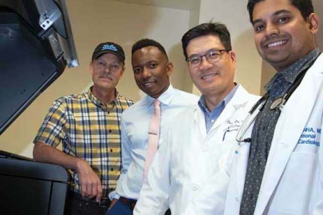
The in-house program at UCLA Health was made possible thanks to a partnership with the University of California, Irvine, and the collaborative efforts of many internal departments. To get the pilot program up and running, Dr. Sinha relied on the expertise of Daniel Levi, MD, and Jamil Aboulhosn, MD, UCLA Health cardiologists and leaders in the field of congenital interventions. Dr. Sinha also tapped the expertise of leadership in the Ahmanson/UCLA Adult Congenital Heart Disease Center, Benjamin Wu, DDS, PhD, director of the UCLA Jane & Jerry Weintraub Center for Reconstructive Biotechnology, and David T. Nelson and Marc-Anthony Lecky from the UCLA Department of Radiology. The technology experts at the Weintraub Center currently oversee the printing of all heart models.
While many medical centers nationwide use these models to plan surgeries, UCLA Health is among a select few centers that can create models in less than 48 hours using on-site technology. “At UCLA Health, we do many cutting-edge procedures to treat congenital and structural heart disease. It’s helpful to be able to visualize how blood vessels interact and how a procedure might affect blood flow. We want our doctors to have every tool available as they prepare for a surgery.” To learn more, visit uclahealth.org/heart.