Key takeaways:
- UCLA researchers show that wildfire smoke attacks two airway defenses at once, paralyzing the cilia that sweep mucus out of the lungs and loosening the protein seals between the cells that line the airway.
- Blocking a cell signaling pathway called MEK/ERK before smoke exposure preserved both cilia movement and the airway barrier in mini human airways grown from stem cells.
- The team is working to develop an inhaler or nasal spray that blocks the pathway — something firefighters and others at high risk could use ahead of a smoky day.
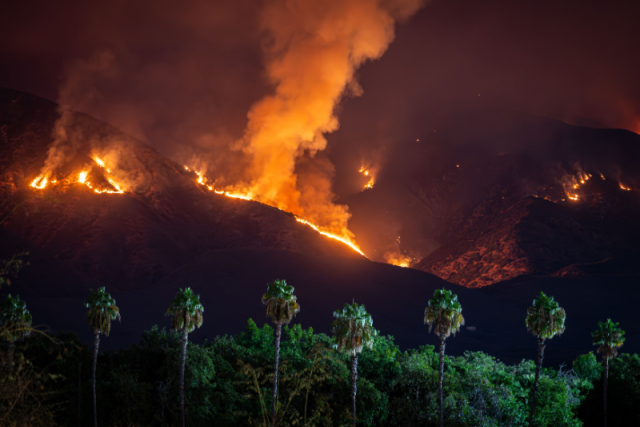
When the Palisades and Eaton wildfires tore through Los Angeles in January 2025, smoke blanketed the region and air quality plunged into the hazardous range. It was a vivid example of a growing problem: After years of improvement, wildfire smoke is pulling air quality back in the wrong direction, and fire season, once confined to hot, dry summers, now stretches across the year.
Fine smoke particles have been linked to lung disease, stroke, heart disease and cancer. But how they injure the cells lining our airways has been difficult to pin down.
Normally, the airway has two lines of defense. Sticky mucus traps inhaled viruses, dust and pollution, and tiny hair-like structures called cilia sweep that mucus up and out. Those cilia grow from cells in the airway lining, which sit packed together like bricks held by cement, forming a wall that keeps whatever the mucus doesn’t catch from reaching the rest of the body.
Now, UCLA researchers have shown that wildfire smoke attacks both defenses at once, paralyzing the cilia and loosening the “cement” between cells. They also identified a cell signal that contributes to the damage and found that blocking it protected human airway tissue in the lab. The findings were published in Environmental Health Perspectives.
“People talk about the lung as a gateway,” said co-senior author Dr. Brigitte Gomperts, a professor of pediatrics and pulmonary medicine at the David Geffen School of Medicine at UCLA and the associate director of translational research at the UCLA Broad Stem Cell Research Center. “If the particles are small enough, and if they can break down the airway barrier, they can get into your brain and your heart, where they can cause inflammation. I call it an insidious threat.”
Evidence from the 2025 Los Angeles wildfires
To see what the smoke did to human airways, the team started with nasal and sinus tissue — part of the upper airway — from 14 Los Angeles residents who had sinus surgery either before the fires, during them, or in the two weeks of smoldering afterward. Each patient was sampled at only one of those points.
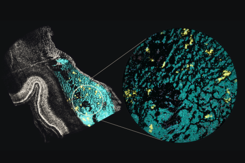
Before the fires, the patients’ airway lining was intact. During the fires, it was patchy and sloughing off, with some areas missing entirely, and the protein seals between cells had fragmented. In the two weeks of smoldering that followed, the lining had partly recovered, though it remained thinner than before the fires and its seals were only partly restored.
Under an electron microscope, tissue collected during the fires also showed dense, particle-like structures inside cells and in the spaces between them. The team identified these structures by their appearance but did not trace them to the smoke.
Everyone in the group had chronic sinus disease, and no one’s individual smoke exposure could be measured, so the samples alone can’t prove that smoke caused the damage. To test that directly, the team turned to lab-grown human airway tissue.
Mini human airways: cilia stall, barrier breaks
First the researchers needed a way to expose that tissue to smoke.
“I came into the lab ready to study wildfire smoke, but without a real system to do so,” said first author Dr. Caroline Cherry, a postdoctoral scholar in the Gomperts lab.
So she improvised, adapting an exposure chamber a labmate had built for cigarette smoke. She ground pine bark from a tree on the UCLA campus, rolled it into tiny “wooden cigarettes” and used a programmable pump to draw the smoke over the tissue. UCLA atmospheric and environmental health scientists found that the smoke’s particle sizes and chemistry were consistent with those of wood smoke measured in the environment.
The tissue itself was grown from airway stem cells isolated from donated lungs with no lung disease. Because these stem cells can renew themselves and mature into the airway’s many cell types, the resulting mini airways contained beating cilia and the protein “cement” between cells, much like the real thing. The team exposed the mini airways to smoke for three minutes a day for five days.
The damage accumulated with each exposure, even though the tissue got clean air in between. In unexposed tissue, about 71% of the surface showed coordinated ciliary movement. After five days of smoke, that fell to about 25%, even though the cells themselves weren’t dying off. The airway’s second defense suffered as well, as the seals between cells loosened and the barrier weakened.
Recovery was only partial. Five days after the final exposure, ciliary movement had climbed back only to about 46%, and the barrier was still weakened. While the cilia are still impaired, the airway can’t clear particles as well.
“It’s concerning in terms of general public health, because that’s quite some time for the particles to be sitting in our lungs,” Cherry said.
Blocking the signal behind the damage
To find out what was contributing to the damage, the team worked with the lab of Dr. Mehdi Bouhaddou, an assistant professor of microbiology, immunology and molecular genetics at the Geffen School of Medicine, to look at protein phosphorylation, the process cells use to switch proteins on and off by adding tiny chemical tags. Smoke scrambled these tags, especially on proteins that build cilia and hold the cell’s internal scaffolding in place.
Different signaling pathways tag different proteins, so the pattern smoke left behind pointed back to the pathways responsible. One stood out among those that control cilia and the cell’s internal scaffolding: MEK/ERK, which also helps regulate cell growth and is already the target of several cancer drugs. Follow-up tests confirmed the pathway was switched on in smoke-exposed tissue.
Live recordings show the movement of tiny hair-like structures called cilia on the surface of human airway cells. In the healthy control (left), cilia beat rapidly and in a coordinated pattern to help clear harmful particles from the lungs. After exposure to wildfire smoke (middle), cilia movement is severely impaired. When cells are treated with a drug that blocks the MEK signaling pathway (right), cilia movement is preserved, demonstrating a potential way to protect the lungs from smoke-related damage. | Credit: Caroline Cherry/Gomperts Lab
So the team tried blocking it, treating the tissue with an MEK inhibitor called pimasertib 30 minutes before each smoke exposure. In this set of experiments, about 48% of the surface showed coordinated ciliary movement in tissue that got no smoke. In tissue exposed to smoke without the drug, that fell to about 14%. At the highest dose of pimasertib, it maintained about 41% ciliary movement. Microscopy showed that the drug kept the cilia’s internal structure intact and helped preserve the seals between cells.
The day the team saw the cilia still beating despite the smoke was “one of the happiest days ever in the lab,” said Gomperts, who is also the co-director of cancer stem cell biology at the UCLA Health Jonsson Comprehensive Cancer Center.
Developing a shield for the airways
Because the drugs were given before each exposure, these experiments tested prevention rather than repair. The team imagines a product that works the same way: an inhaler or nasal spray taken ahead of a smoky day, delivering protection straight to the airway.
“This would be for firefighters and the military population who are exposed to burn pits,” Cherry said. “But we also see this for those more at risk of having severe consequences from smoke exposure, like kids and people with lung disease or heart disease.”
This is early-stage work. Next steps include formulating the compound for inhalation and conducting safety studies.
“Our goal is to develop a product that’s affordable and available over the counter,” Gomperts said. “We want anyone around the world to be able to get it.”
The compositions and methods described in this study are covered by a patent application filed by the UCLA Technology Development Group on behalf of the Regents of the University of California, with Gomperts and Cherry listed as inventors.
Co-senior authors are Mehdi Bouhaddou of UCLA and Amarbir Gill of the University of Michigan. Additional authors include Declan Winters, Abdo Durra, Chandani Sen, Andrew Frauenpreis, Emily Hou, Coline Luo, Tammy Rickabaugh, Luca Lit, Sarah Siegel, Matthew Aaron Blumberg, Kristen Castillo, Andrew Lund, Catherine Banach, Zhuoying Lin, Saagar Patel, Mikayla Murphree, Sara Makanani, Prashant Kaushal, Immy Ashley, Jeffrey Suh, Nicholas Jackson, David Gonzalez, Yifang Zhu, Suzanne Paulson, Chen Yuan Kam, Daniel Beswick and Eszter Vladar.
This research was supported by the University of California Office of the President’s California Firefighter Cancer Prevention and Research Program, the National Institutes of Health, the UCLA Environmental and Molecular Toxicology Program, the Tobacco-Related Disease Research Program, the Burroughs Wellcome Fund, the UCLA Health Jonsson Comprehensive Cancer Center, the Howard Hughes Medical Institute, the UCLA Molecular Biology Institute, the UCLA Cellular and Molecular Biology Training Program, the UCLA Health Jonsson Comprehensive Cancer Center and UCLA Broad Stem Cell Research Center Ablon Scholars Program, the UCLA AIDS Institute, the James B. Pendleton Charitable Trust and the McCarthy Family Foundation.