Imaging Tests
Imaging Studies for patients with Polycystic Kidney Disease (PKD) on Hemodialysis
The diagnosis of ADPKD varies depending on the stage of the disease. Once the disease has fully developed, diagnosis relies on the clinical assessment involving the patient's medical history and physical examination. However, establishing a definite diagnosis can be challenging due to the presence of similar symptoms in other disorders. Therefore, additional methods such as diagnostic imaging and genetic testing play a crucial role in confirming the diagnosis.
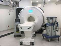
Imaging technologies like ultrasound, computed axial tomography, and nuclear magnetic resonance are employed to identify cysts in the kidneys, liver, or pancreas. Magnetic resonance imaging has demonstrated higher sensitivity to ultrasound, enabling measurements like height-adjusted total kidney volume and providing clearer cyst definition without the need for contrast agents.
Yet, these imaging procedures come at a significant cost and are often omitted for certain subsets of the ADPKD population, such as young individuals at risk or patients with atypical or newly occurring renal cystic disease. In these cases, genetic testing serves as the definitive approach.
Criteria for Diagnosing PKD:
Current screening criteria states:
- Two or more cysts as diagnostic for individuals ages less than 59
- A minimum of four cysts in each kidney for older at-risk individuals.
The finding of fewer than two cysts in each kidney is sufficient to exclude the disease in people who are 30 or older.
Disclaimer: The UCLA Health System cannot guarantee the accuracy of such information. The information is provided without warranty or guarantee of any kind. Please speak to your Physician before making any changes.