X-Ray / Fluoro: Radiography vs. Fluoroscopy
Find Your Care
Radiologists are experts in all types of imaging, including advanced techniques.
Call today to find medical imaging near you and schedule your imaging procedure (MR, CT, PET, Dexa, Ultrasound).
Radiography is the grandfather of human imaging studies. Revolutionary refinements have taken place since 1895 when Wilhelm Roentgen first made an image of his wife's hand and wedding ring.

What is radiography
During a radiographic procedure, an X-ray beam is passed through the body. A portion of the X-rays are absorbed or scattered by the internal structure and the remaining X-ray pattern is transmitted to a detector so that an image may be recorded for later evaluation.
The modern radiograph is usually a computerized image. It is the state-of-the-art technique to look for community acquired pneumonia and congestive heart failure. Fractures and arthritis are also commonly well imaged by radiography.
What is fluoroscopy
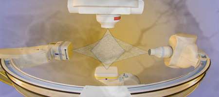
When the X-ray beam is used with a video screen, the technique is called fluoroscopy. This allows physicians to visualize the movement of a body part or of an instrument or dye (contrast agent) through the body in real time.
Fluoroscopy studies such as the upper gastrointestinal series are popular to evaluate patients with suspected gastroesophageal reflux and other problems such as swallowing difficulty.