• UCLA Health
  • myUCLAhealth
  • School of Medicine
Radiation Oncology UCLA

UCLA Radiation Oncology

Radiation Oncology UCLA
  • About Us
    • Chair's Welcome
    • Request an Appointment
    • Refer a Patient
    • Contact Us
    • How You Can Help
  • Our Team
  • Patients & Treatments
    • First Visit for Patients
    • Patient Education Videos
    • FAQs
    • Patient Stories
    • Testimonials
    • Treatments
    • Resources / Links
    • Clinical Trials
    • Request an Appointment
    • Health Plans
    • Contact Us
  • Research & Technologies
    • Innovation & Technology
    • Cutting-Edge Devices
    • Division of Molecular and Cellular Oncology
    • Medical Physics Research
    • Research Labs
    • Scholarly Activity
    • Translational Research
    • Clinical Trials
    • PSMA Risk Calculator
  • Education & Training
    • Welcome and Overview
    • Residency and Fellowship Recruitment
    • Medical Residency
    • Residents
    • Medical Physics Residency Program
    • Medical Student Rotation
    • Brachytherapy Fellowship
    • Medical Student Preceptorship Program
    • BioMedical Physics Graduate Program
  • News & Events
    • Conferences & Courses
  • UCLA Health
  • myUCLAhealth
  • School of Medicine

UCLA Radiation Oncology

External Beam Radiation Therapy (EBRT)

  1. Home
  2. Research & Technologies
  3. Innovation & Technology
  4. External Beam Radiation Therapy (EBRT)

External Beam Radiation Therapy (EBRT)

3D conformal therapy
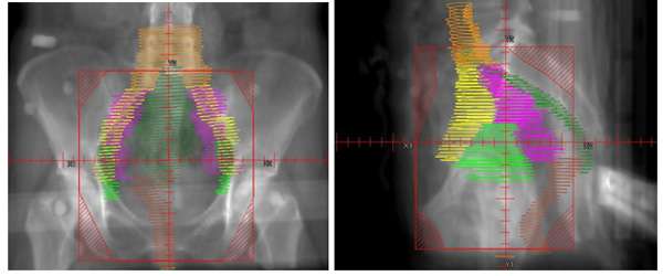
This is a radiation technique where multiple radiation beams are used to conform to a target. The dose of radiation that is delivered from each radiation field is shaped by small leaves in the head of the radiation treatment machine called multileaf collimators. The intensity of the radiation from each field is uniform which is different than with intensity modulated radiation therapy (IMRT). Below is an example of what the treatment fields look like on an X-ray when treating the pelvis.

X-ray Film

*Images from wikibooks -- In the image on the left, the red outline depicts the borders of the radiation field on this “AP” (anterior-posterior) X-ray film. The striped lines in the corners represent areas where the radiation is blocked from penetrating. The colored structures in the middle of the field represent contours of normal organs and the areas being targeted for radiation. The image on the right represents a representative pelvic field on a lateral X-ray. Below is an example of the distribution of the radiation dose on an axial CT scan using this type of “4-field box” approach.


X-ray Film

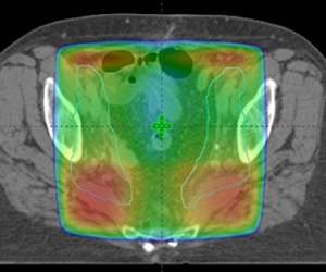
One can see that the radiation dose covers the entire pelvic region but the actual target on this slice, which can be seen in the pale blue outlines on the right and left sides of the pelvis, is actually much smaller. Ideally we would be able to sculpt the radiation to these pale blue regions and to spare the middle part of the pelvis from unnecessary radiation. Advances in radiation with intensity modulated radiation therapy (IMRT) give us the ability to accomplish this.

  • Request an appointment >
  • Westwood: 310-825-9775
  • Santa Monica: 424-259-8777

In this Section

  • Intensity Modulated Radiotherapy (IMRT)
  • Stereotactic Radiosurgery (SRS)
  • Stereotactic Body Radiation Therapy (SBRT)
Facebook Twitter Instagram Youtube LinkedIn
UCLA Health hospitals ranked best hospitals by U.S. News & World Report
  • UCLA Health
  • Find a Doctor
  • School of Medicine
  • School of Nursing
  • UCLA Campus
  • Directory
  • Newsroom
  • Subscribe
  • Patient Stories
  • Giving
  • Careers
  • Volunteer
  • International Services
  • Privacy Practices
  • Nondiscrimination
  • Billing
  • Health Plans
  • Emergency
  • Report Broken Links
  • Terms of Use
  • 310-825-9775
  • Maps & Directions
  • Contact Us
  • Your Feedback
  • Report Misconduct
  • Get Social
  • Sitemap