Endocrine Surgery
Abdominal Imaging for Adrenal Tumors
Find Your Care
We deliver effective, minimally invasive treatments in a caring environment. Call 310-267-7838 to connect with an expert in endocrine surgery.
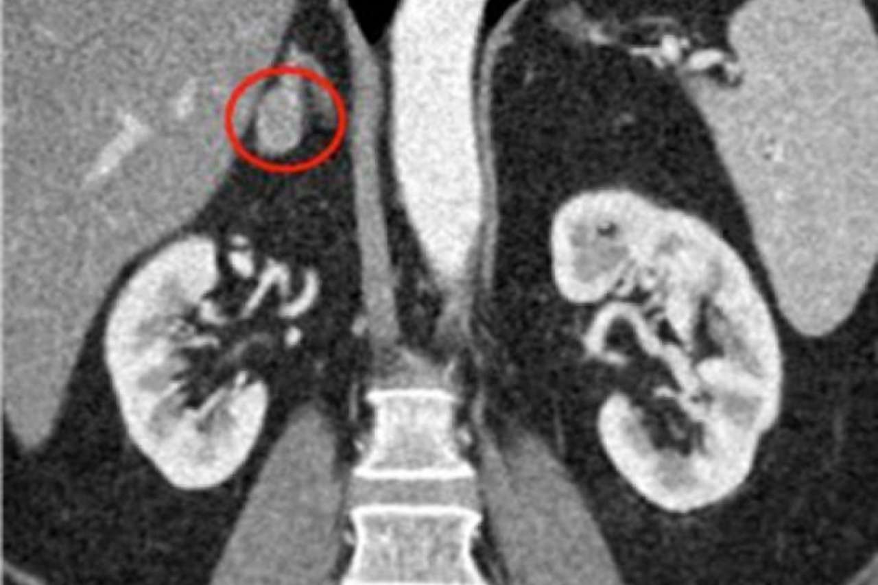
Adrenal CT or MRI are the most common ways to identify adrenal tumors.
Imaging characteristics of benign adrenal nodules:
- Well-Defined Borders: Benign nodules usually have clear and well-defined borders on imaging scans, making it easier to distinguish them from surrounding tissues.
- Low density / lipid rich: Benign nodules are often more fatty and less dense than the surrounding structures.
- Smooth and Regular Shape: They often appear round or oval with a smooth and regular shape. This contrasts with irregular shapes that might be seen in more worrisome nodules.
- Consistent Density: Benign nodules tend to have a consistent density on imaging, indicating that the tissue is uniform throughout and doesn't show signs of abnormal growth.
- Stable Size Over Time: In many cases, benign nodules remain the same size over time. Monitoring their size through follow-up imaging can help confirm their non-cancerous nature.
- Non-Invasive Appearance: They typically do not invade surrounding tissues or structures. This lack of invasion is a positive sign in distinguishing benign nodules from potentially harmful ones.
Adrenal tumors that are larger than 4 cm in size or are enlarging over time often need to be removed due to an increased risk of malignancy.
Red circles show small adrenal tumors.
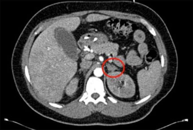
Red circles show small adrenal tumors.