• UCLA Health
  • myUCLAhealth
  • School of Medicine
Pediatric Neurosurgery

Pediatric Neurosurgery

Pediatric Neurosurgery
  • About Us
    • Family-Centered Care
    • Chase Child Life
    • In the News
    • Webinars
  • Meet Our Team
  • Conditions & Treatment
    • Pediatric Brain Tumor Program
    • Pediatric Chiari and Syringomyelia Program
    • Pediatric Concussion Program
    • Pediatric Craniosynostosis Program
    • Pediatric Epilepsy Surgery
    • Pediatric Hydrocephalus Program
    • Pediatric Neurovascular Program
    • Pediatric Scoliosis Program
  • Resources for Families
    • A Guide to Preparing for Surgery
    • Admission Information
    • Facilities & Amenities
    • Visitors
    • Medical Records
    • UCLA Neurosurgery App
  • Giving
  • Patient Stories
    • Rhianna's Story - Chiari Malformation
    • Cameron’s Story - Brain Tumor
    • Kobe's Story - Using Virtual Reality for Brain Surgery
    • Brock's Story - Brain Seizures
    • Nathan's Story - Tuberous Sclerosis Complex
    • Nikki's Story - Epilepsy
    • Aiden's Story - Sturge-Weber Syndrome
    • Lucy's Story - Tuberous Sclerosis
    • Nichele - Spinabifida
  • Contact Us
    • Appointment Request
    • Telemedicine and Remote Second Opinion
    • Maps and Directions
  • UCLA Health
  • myUCLAhealth
  • School of Medicine

Pediatric Neurosurgery

Conditions & Treatment

Conditions & Treatment

Conditions & Treatment

  • Pediatric Brain Tumor Program
    • Brain Tumors
    • Our Physician Team
    • Referring Physicians
  • Pediatric Chiari and Syringomyelia Program
    • Faculty
    • Frequently Asked Questions
  • Pediatric Concussion Program
  • Pediatric Craniosynostosis Program
  • Pediatric Epilepsy Surgery
    • Epilepsy FAQs
    • Diseases and Conditions
      • Focal Cortical Dysplasia
      • Hemimegalencephaly
      • Rasmussen’s Encephalitis
      • Sturge-Weber Syndrome
    • Diagnostic Operations
    • Epilepsy Treatment
      • Hemispherectomy
      • Laser Interstitial Thermal Therapy
    • Our Epilepsy Team
    • Patient Services
    • Patient Stories
    • Helpful Links
    • Hemispherectomy Outcome Prediction Scale (HOPS)
    • High Frequency Oscillation
    • Rare Epilepsies & Brain Disease Tissue Bank (REBDTB)
      • Participate in Ongoing Research
      • Donate Biological Samples To The Tissue Bank
      • Submit a Proposal For Research
      • About the Rare Epilepsies & Brain Disease Tissue Bank
      • Contact the Rare Epilepsies & Brain Disease Tissue Bank
  • Pediatric Hydrocephalus Program
    • Hydrocephalus FAQs
    • Faculty
  • Pediatric Neurovascular Program
  • Pediatric Scoliosis Program
  • Pediatric Brain Tumor Program
  • Pediatric Chiari and Syringomyelia Program
  • Pediatric Concussion Program
  • Pediatric Craniosynostosis Program
  • Pediatric Epilepsy Surgery
  • Pediatric Hydrocephalus Program
  • Pediatric Neurovascular Program
  • Pediatric Scoliosis Program
  • Epilepsy FAQs
  • Diseases and Conditions
  • Diagnostic Operations
  • Epilepsy Treatment
  • Our Epilepsy Team
  • Patient Services
  • Patient Stories
  • Helpful Links
  • Hemispherectomy Outcome Prediction Scale (HOPS)
  • High Frequency Oscillation
  • Rare Epilepsies & Brain Disease Tissue Bank (REBDTB)
  • Focal Cortical Dysplasia
  • Hemimegalencephaly
  • Rasmussen’s Encephalitis
  • Sturge-Weber Syndrome
  1. Home
  2. Conditions & Treatment
  3. Pediatric Epilepsy Surgery
  4. Diseases and Conditions
  5. Focal Cortical Dysplasia

Focal Cortical Dysplasia

Share this

Special Consideration: Epilepsy Surgery for Focal Cortical Dysplasia

In this section: Tuberous Sclerosis Complex | Focal Cortical Dysplasia | Hemimegalencephaly | Rasmussen’s Encephalitis | Sturge-Weber Syndrome

Epilepsy surgery for Focal Cortical Dysplasia at UCLA
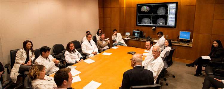
Focal Cortical Dysplasia Team at UCLA

Focal cortical dysplasia is a congenital abnormality where there is abnormal organization of the layers of the brain and bizarre appearing neurons. There are both genetic and acquired factors that are involved in the development of cortical dysplasia.  In general, there are three pathological subtypes of cortical dysplasia that are recognized.  These lesions have a high propensity to cause epilepsy that do not respond to medications. They are in fact the most common reason to require an epilepsy operation in children.

Surgical Treatment of Epilepsy in Children with Focal Cortical Dysplasia

Occasionally, focal cortical dysplasia can be highly difficult to detect or may remain invisible on MRI.  Other times, the affected area of the brain can be larger than the abnormality revealed by the MRI, which can be a possible cause of poor outcomes if surgery is based on the MRI data alone.  For this reason, several other imaging modalities are very helpful to help delineate the abnormality. Dr. Noriko Salamon from the Department of Neuroradiology uses advanced imaging techniques through the fusion of a high resolution MRI with an FDG-PET study, a UCLA invention, to detect these subtle lesions.  FDG-PET can demonstrate the area of reduced metabolism affected by the dysplasia.  Magnetoencephalography is a non-invasive technique that can localize the abnormal electrical activity to find and assess the size of the seizure focus.  Diffusion tensor imaging analyzes the free water molecules in the brain to help identify and track abnormal fibers of the brain that may be associated with cortical dysplasia.  Sometimes, invasive techniques such as an intracranial EEG study may be required to accurately map the seizure onset in relation to the focal cortical dysplasia.  These technique gives surgical treatment options to patients who would otherwise not be candidates for a curative procedure.

Resective surgical options which are aimed at stopping seizures altogether include lesionectomies, lobectomies and in certain cases, hemispherectomies.   Generally, the younger the child or infant, the larger an operation is required.  This is because focal cortical dysplasia type II is most commonly found in very young children and the abnormality is more extensive.  In contrast, surgeries in older children and young adults tend to be for focal cortical dysplasia type I, and is characterized by less extensive abnormalities most commonly found in the temporal lobe.

Following surgery, 60-80% of children remain seizure-free, depending on the center at which this is performed.  Some of the most favorable factors that predict a successful surgery include a total resection of the focal cortical dysplasia.  Sometimes, full resection of the abnormality may not be deemed suitable if it involves resection of a neurologically important structure (such as motor, sensory or speech related brain tissue).  In these situations, newer surgical techniques have been developed that may be an option.

Surgery carries risks related to infection, blood transfusion (commonly in infants), and specific risks related to the brain tissue that needs to be removed.  Permanent deficits such as new neurological deficits or hydrocephalus are rare.  Seizure freedom is determined generally after at least 1 year and sometimes 2 years following surgery.  If surgery is successful, there is a 50% chance of being able to stop medications completely.  Other benefits may include improvement in behavior, concentration, attention, cognition and development.

< Return to Diseases and Conditions

Like Us on Facebook Follow Us on Twitter Subscribe to Our Videos on YouTube Follow us on Instagram Connect with Us on LinkedIn Follow us on Pinterest
  • UCLA Health
  • Find a Doctor
  • School of Medicine
  • School of Nursing
  • UCLA Campus
  • Directory
  • Newsroom
  • Subscribe
  • Patient Stories
  • Giving
  • Careers
  • Volunteer
  • International Services
  • Privacy Practices
  • Nondiscrimination
  • Billing
  • Health Plans
  • Emergency
  • Report Broken Links
  • Terms of Use
  • 1-310-825-2631
  • Maps & Directions
  • Contact Us
  • Your Feedback
  • Report Misconduct
  • Get Social
  • Sitemap
Like Us on Facebook Follow Us on Twitter Subscribe to Our Videos on YouTube Follow us on Instagram Connect with Us on LinkedIn Follow us on Pinterest

Sign in to myUCLAhealth

Learn more about myUCLAhealth