History of BIRC Research
The Research History Behind BIRC
Acutely After Traumatic Brain Injury
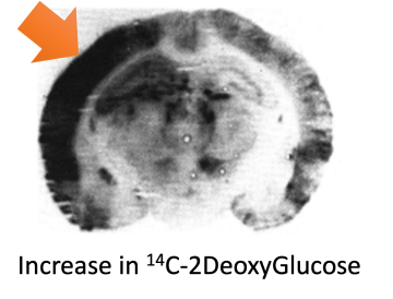
In 1990, Dr. David Hovda and Team demonstrated that Traumatic Brain Injury resulted in early increase in glucose uptake into the brain and that this uptake was dependent on excitatory amino acid receptors and involved calcium uptake (Hovda et al., 1990).
Subsequent work elaborated on this initial discovery to show that Acute increase in glucose uptake was initiated by neuronal discharge and subsequent activation of excitatory amino acid receptors (Katayama et al., 1990; Kawamata 1992; Hovda 1991).
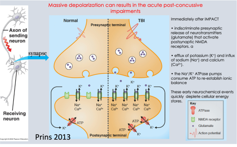
We now know that the impact of TBI causes the brain to move and brain cells release their signaling chemicals.
- These bind to the receptors on the other side and open up channels where ions like potassium, sodium and calcium flux down their concentration gradients.
- This action causes the pumps to be activated, which increases the need for energy!
Prolonged Metabolic Depression After Traumatic Brain Injury
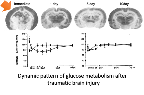
The time course of the brain’s glucose metabolic disruption after TBI was determined in 1991 (Yoshino et al., 1991). This was the first paper to show that the immediate increase in glucose metabolism was followed by a prolonged depression.
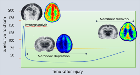
In 1995, the concussion pathophysiology image that is frequently used to describe the time course of changes after TBI was created by Mayumi Prins and published in Hovda et al., 1995.
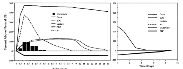
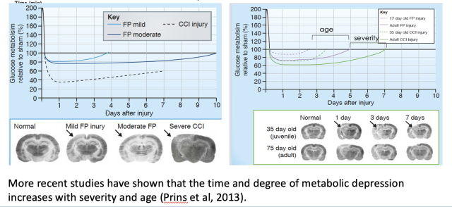
This dynamic pattern of brain glucose metabolism has been repeatedly demonstrated in preclinical models of TBI and in human TBI subject (Bergsneider et al., 1997).
THIS is the research on which the UCLA BRAIN INJURY RESEARCH CENTER WAS FOUNDED.