Biochemistry & Molecular Biology Instrumentation Core (BMBI)
The Biochemistry & Molecular Biology Instrumentation (BMBI) module is an essential core resource to support the preparation and analysis of biological samples (RNA, DNA, and protein). Researchers are encouraged to seek assistance from the core director (Dr. Radu) or staff research associates to select the appropriate instruments for specific analysis, to schedule & properly use the instruments, and to obtain protocols to prepare the samples. Instrument/equipment is listed under three categories:
I.Basic instrumentations (centrifuges/rotors; incubators; freezers; PCR; etc)
II. Specialized spectrophotometers and protein dynamic analyzers
III. Highly-sensitive instruments for protein quantification and biochemical assays
Trained and authorized personnel may make reservations to use the facilities at https://uclajsei.bookmylab.com/
I. Basic instrumentations (centrifuges, incubators, cyclers, gel imagers, etc.)
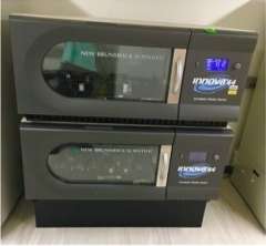
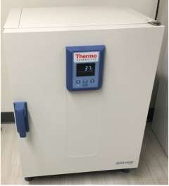
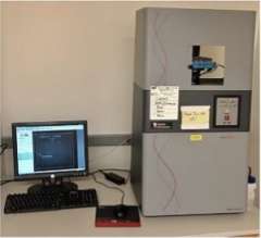
The BMBI core module resources consist of common instrumentations such as autoclaves, ultra-low temperature freezers, various centrifuges, incubators, an Alpha Innotech multi-imaging instrument, polymerase chain reaction (PCR) machines for DNA and RNA analysis (Bio-Rad Dual C1000 Thermal Cycler). Investigators can also determine gene expression profiles using high throughput microfluidic assays (BioMark Fluidigm Thermal Cycler), a next generation single cell RNA-sequencing (scRNA-seq) PCR approach.
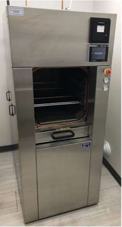
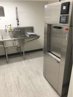
Beta Star Autoclaves
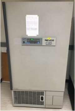
Ultra-low Temperature Freezers

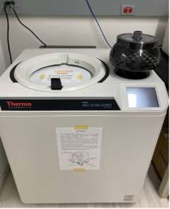
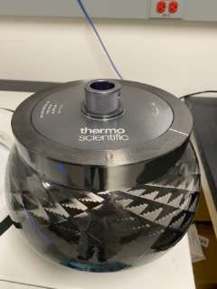
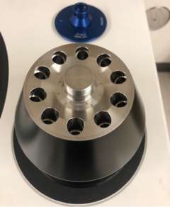
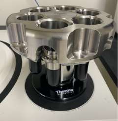
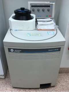
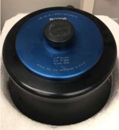
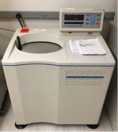
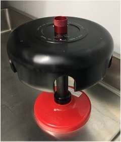
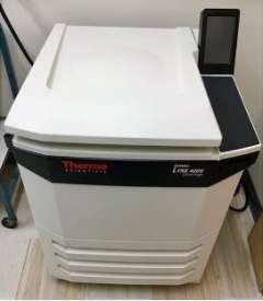
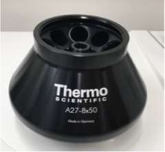
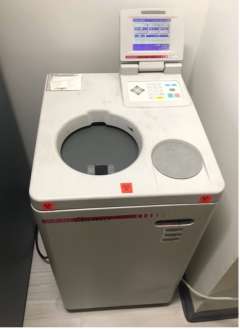
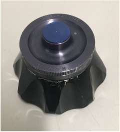
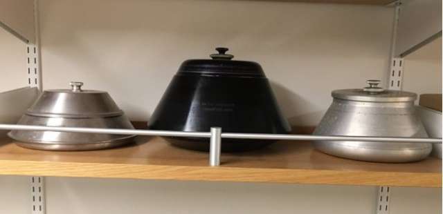
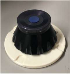
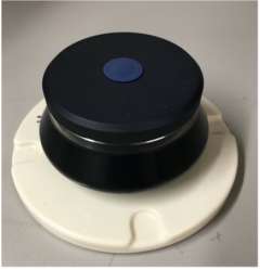
Centrifuges and Rotors
Max speed: 80,000 rpm
Temperature range: 0 to 40°C
Max speed: 50,000 rpm
Capacity: 8 x 39 ml, angle: 23°
Max speed: 80,000 rpm
Capacity: 8 x 4.4 mL, angle: 25°
Max speed: 30,000 rpm
Capacity: 6 x 39 mL, angle: 90°
Max speed: 26,000 rpm
Temperature range: –10 to 40°C
Max speed: 25,000 rpm
Capacity: 8 x 50 mL, angle: 34°
Max speed: 28,000 rpm
Temperature range: 0 to 40°C.
Max speed: 80,000 rpm
Capacity: 6 x 38.50 mL, angle: 90°
Max speed: 24,000 rpm
Temperature range: –10 to 40°C
Max speed: 27,000 rpm
Capacity: 8 x 50 ml, angle: 34°
Max speed: 150,000 rpm
Temperature range: 0 to 40°C
Max speed: 150,000 rpm
Capacity: 8 x 2 mL, angle 30°
Other Rotors:
Sorvall SA-300 fixed angle rotor
Max speed: 25,000 rpm, capacity: 6 x 50 mL, angle: 34°.
Sorvall M-24 fixed angle rotor
Max speed: 20,500 rpm, capacity: 24 x 16 mL, angle: 28°.
Sorvall GSA fixed angle rotor
Max speed: 13,000 rpm, capacity: 6 x 250 mL, angle: 28°.
Sorvall HS-4 swinging-bucket rotor
Max speed: 7,500 rpm, capacity: 4 x 250 mL, angle: 90°.
- Features a CCD camera, transillumination and reflective imaging modes, and UV and visible light sources. Filters for imaging Ethidium Bromide, Cy2, Cy3, Cy5, and Chemiluminescence.
- Blue, green, and red LEDs and filters for acquisition of single color and multicolor fluorescence images.
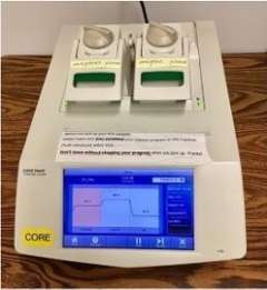
- Two 48-well modules with adjustable heated lids to support a wide range of vessels, including low-profile and standard-height PCR plates.
- Two protocol programming options, including Protocol Autowriter, which automatically generates customized standard, fast, and ultrafast protocols.
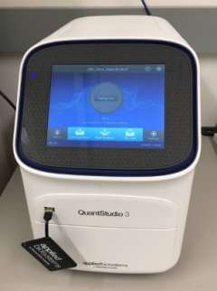
- Real-Time PCR system can detect very small changes, as low as 1.5 fold, in gene expression, that may be missed by other systems.
- Dynamic range of up to 10 logs tracks broad changes in expression between different types of samples and conditions.
- Excitation filters: 470 ± 15nm, 520 ± 10nm, 580 ± 10nm, 640 ± 10 nm
- Emission filters: 520 ± 15nm, 558 ± 12 nm, 586 ± 10 nm, 623 ± 14 nm
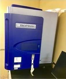
- Supports multiple integrated fluid circuit (IFC) formats for gene expression, genotyping, and digital PCR.
- Processes up to 192 samples per run.
- Detection: CCD camera; 9M pixel, 12 μm x 12 μm pixel size.
- Fluorescence excitation: 475 nm, 530 nm, 575 nm.
- Fluorescence emission: 525 nm, 570 nm, 630 nm.
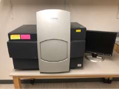
II. Specialized spectrophotometers and protein dynamic analyzers
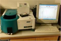
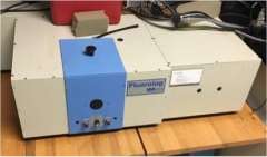
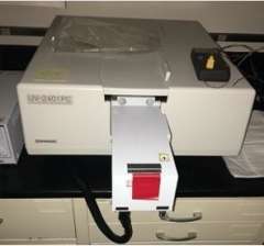
Besides other common instruments (Shimadzu UV-2600 and UV-2700 spectrophotometers), microplate readers (Molecular Devices SpectraMax iD3), immunoassay protein analyzers, this module has available specialized spectrometers for detailed protein structural and functional analysis, including fluorescence, UV-visible absorbance (Agilent Cary Eclipse and Horiba Fluorolog spectrophotometer), Fourier transform infra-red, and customized circular dichroism (CD) spectroscopies and sophisticated multi-angle and quasi-elastic light scattering instruments are also available (Bruker Tensor spectrometer).
- Fluorescence spectroscopy with multiple data collection modes including fluorescence, phosphorescence, chemiluminescence, and bioluminescence.
- Features a xenon flash lamp, captures a data point every 12.5 ms, and scans at 24,000 nm/min without peak shifts.
- Accessories include temperature control and solid sample handling, automatic polarizers and microplate reader for high sample throughput.

- Fourier Transform Infrared (FT-IR) spectrometer for analyzing compounds.
- Spectral Range: 7,500 to 370 cm-1, with standard KBr beamsplitter.
- Resolution: Better than 1 cm-1 (apodized).
- Scan Speed: 3 velocities, 2.2 - 20 kHz (1.4 - 12.7 mm/sec opd).
- For analysis of steady-state or molecular dynamics as well as IR probes.
- Can detect 50-femtomolar fluorescein.
- Features all-reflective optics for high sensitivity at all wavelengths and for microsamples.
- Wavelength Accuracy ± 0.5 nm. Integration Time: 1ms – 160s.
- Single monochromator with a high-performance blazed holographic grating in the aberration corrected Czerny Turner mounting.
- Wavelength range: 190 to 900nm (performance guaranteed range). Extendable to 1,100nm with optional photomultiplier.
- Wavelength repeatability: ±0.1nm. Wavelength accuracy: ±0.3nm.
- Resolution: 0.1nm. Spectral bandwidth: 0.1, 0.2, 0.5, 1, 2 and 5nm.
III. Highly-sensitive instruments for protein quantification and biochemical assays
Researchers have access to an automated Western immunoblot system allowing for quantification of low abundant proteins via a sensitive immunoassay reaction involving minimal sample manipulation with data being analyzed digitally (Automated Protein Simple WES). This acquisition complements two infrared imagers (Li-COR Odyssey CLx) and two NanoDrop ND-1000 spectrophotometers. Proteomics discovery and signal-pathway profiling can be done using our Veritas Laser Capture Microdissection instrument. Additionally, the newly acquired Molecular Devices SpectraMax iD3 microplate reader permits quantification at multiplex formats (6 to 384-well) for absorbance, fluorescence, and luminescence, thus enabling multiple biochemical assays at the same time. Lonza 4D Electroporator was recently received to allow investigators to achive high transfection efficiency directly to nucleus even for non-dividing cells.
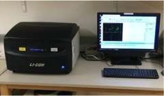
- Features near-infrared (NIR) fluorescence to analyze Western blots, cell-based assays, protein arrays, gel shift assays, and tissue sections.
- System can analyze and quantify several Western blots in a single scan and two different antibodies can be used simultaneously.
- Two solid-state diode lasers at 685nm and 785nm.
- Scanning Speed: 5-40 cm/s. Resolution: 21-337 µm.
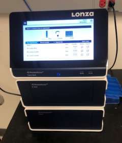
- Is a state-of-the-art electroporation system with capability of optimizing the electrical parameters for specific cell types.
- Transfer can be targeted directly into the cell nucleus, which allows for efficient transfection of even non-dividing cells (such as those found in the retina), resulting in prompt expression from the transfected construct.
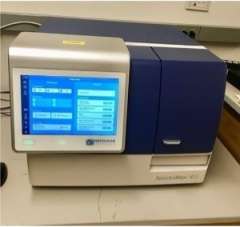
- Reads absorbance, fluorescence, and luminescence in plates with 6 to 384 wells.
- Read types: endpoint, kinetic, spectrum scan, and well scan.
- Wavelength ranges: Abs: 230 - 1000 nm, FL Ex: 250 - 830 nm, FL Em: 270 - 850 nm, Lumi: 300 - 850 nm.
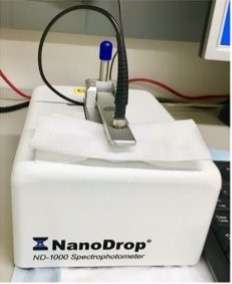
- Full-spectrum spectrophotometer that measures 1 ul samples with high accuracy and reproducibility.
- Path lengths of 1mm and 0.2mm for each measurement cycle provide an extensive dynamic range.
- Wavelength range: 220 to 750 nm. Wavelength accuracy: 1 nm. Wavelength resolution: 3 nm.
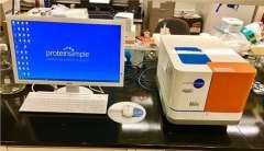
- Wes separates and analyzes proteins by size from 2-440 kDa, and runs up to 25 samples in 3 hours.
- Simply load your samples and reagents into an assay plate, and Wes automates all steps including sample loading, size-based protein separation, immunoprobing or total protein labeling, washing, detection, and data analysis.
- Sample required: 0.3-1.2 µg (protein), 0.6-1.2 µg (immunoassay). Volume required: 3 µL/well.
- Cutting-edge technology to procure cells/regions of interest from tissues under direct microscopic visualization.
- LCM enables analyzing specific cell populations within the context of their heterogeneous tissue microecology.
- A variety of downstream applications: DNA genotyping, RNA transcription, cDNA library generation, proteomics discovery and signal-pathway profiling.