Microscopy and Imaging Analysis Core
The M&I core consists of four divisions
- General microscopy, including scanning laser confocal microscopy
- Super-resolution and high-speed microscopy
- Electron microscopy
- Image Analysis
To use these facilities, please contact the core director (Dr Williams) or the appropriate staff technician for authorization and any required training. Trained and authorized personnel may make reservations to use the facilities at https://uclajsei.bookmylab.com/
In addition to instrument training, researchers may seek assistance or guidance with sample preparation, experimental design, and image acquisition.
View Examples of Images and Videos
General microscopy, including scanning laser confocal microscopy
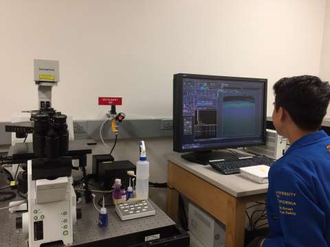
Upright Confocal Microscope - Olympus FluoView FV1000
For imaging fixed or live specimens. Features 4 fluorescence detectors, 1 transmission detector for brightfield or DIC imaging, motorized stage for mosaic stitched imaging, second scan head for FRAP imaging, and capable of XYZT and lambda scanning.
Laser lines: 405, 458, 488, 515, 559, and 635 nm.
Objectives: 10x, 20x, 40x oil, 60x oil, 60x water, and 100x oil.
Inverted Confocal Microscope - Olympus FluoView FV1000
For imaging fixed or live specimens. Features 3 fluorescence detectors, 1 transmission detector for brightfield or DIC imaging, manual stage, and capable of XYZT scanning.
Laser lines: 405, 458, 488, 515, 559, and 635 nm.
Objectives: 10x, 20x, 40x oil, 60x oil, 100x oil.
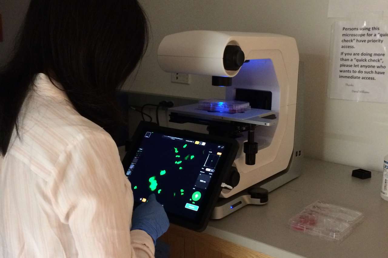
Fluorescence Microscope - Echo Revolve R4
For imaging fixed or live specimens. Features inverted or upright configuration, 13” retina touch display, 3 imaging modes (fluorescence, phase contrast, and brightfield), 3.2mp CMOS monochrome camera for fluorescence/phase contrast, and 12mp CMOS color camera for brightfield.
Filters: DAPI, GFP, and TX RED.
Objectives: 4x, 10x, and 20x.
Dissection Microscope - Leica MZFLIII
For imaging live specimens. Features upright configuration, fluorescence and brightfield imaging, 10x eyepieces, 10:1 zoom, and 5mp CCD camera.
Filters: GFP and TX RED.
Objective: 1x.
Light Microscope - Zeiss Axiophot
For imaging fixed specimens. Features upright configuration, brightfield and phase contrast imaging, Photometrics Coolsnap color camera, and Image-Pro Plus software.
Objectives: 2.5x, 5x, 10x, 20x, 40x oil, 63x oil, and 100x oil.
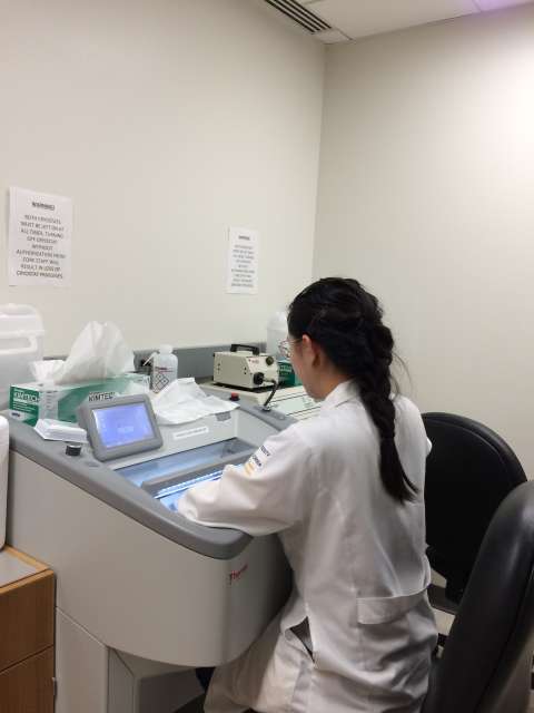
Thermo Scientific and Leica Cryostats
For thick sectioning, typically for immunofluorescence.
Super-resolution and high-speed microscopy
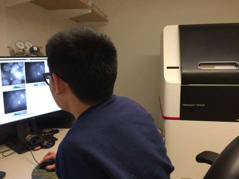
DeltaVision OMX Super-resolution Microscope with Structured Illumination (2D/3D)
- Enhanced resolution in X, Y, and Z axes
- Co-localization using up to 4 fluorescent labeling proteins in live and fixed cells
- Photokinetics with the PK targeting accuracy of 0.5 mm for analysis of interactions between molecules in live cells.
- Ring TIRF (Total Internal Reflection Fluorescence) operation with even illumination.
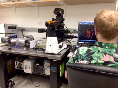
Nikon Eclipse Ti2 Microscope, Yokogawa CSU-22 Spinning Disk Confocal Microscope
25-mm Field of View (FOV), dual EMCCD cameras, delivers high speed image acquisition. This instrument is particularly useful for imaging rapid events in live cells.
Electron microscopy
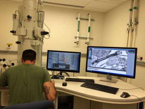
JEOL JEM 1400-Plus transmission electron microscope (TEM)
- Provides up to 120 kV imaging.
- Samples may be imaged in a tilt series for electron tomography, and subsequent 3D reconstruction.
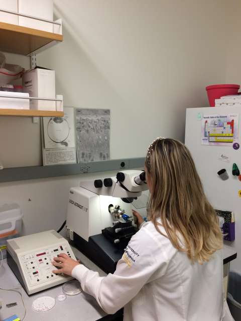
Leica Ultracut UCT and an RMC Powertome X Ultramicrotomes
- Cutting ultrathin sections for TEM, semithin sections for light microscopy, and sections for Intermediate Voltage EM Tomography on instruments at the Electron Imaging Center for NanoMachines (EICN)
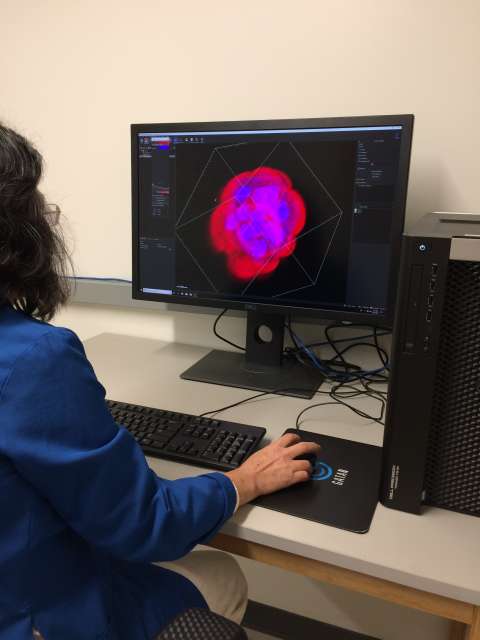
Dell Precision T7910 Workstation: Windows 10, 64GB RAM, and 4GB graphics card.
Software: Imaris, Volocity, FluoView, Image Studio, ImageJ / FIJI.