• UCLA Health
  • myUCLAhealth
  • School of Medicine
UCLA Neurosurgery

UCLA Neurosurgery

UCLA Neurosurgery
  • About Us
    • Chair's Welcome
    • Neurosurgery Faculty
    • Our History
    • Clinical Quality Program
    • Innovation and Technology
    • Giving
    • eNewsletters
    • In the News
    • Webcasts
    • Webinars
    • Resources and Links
    • Global Neuro Health
    • Golden Portal Awards
    • Visionary Ball
    • Lab Party
    • UCLA Neurosurgery App
    • Spotlights
    • Chair's Welcome
    • Neurosurgery Faculty
    • Our History
    • Clinical Quality Program
    • eNewsletters
    • Innovation and Technology
    • In the News
    • Webcasts
    • Webinars
    • Giving
    • Global Neuro Health
    • Lab Party
    • Spotlights
    • Submit Anonymous Feedback (For Faculty)
  • Conditions Treated
    • Alphabetical Conditions
    • Brain Conditions
    • Brain Tumor
    • Pediatric Conditions
    • Spine Conditions
    • Vascular Conditions
    • Acoustic Neuroma
    • Adult Tethered Cord
    • Acromegaly
    • Acute Subdural Hematomas
    • Anaplastic Astrocytoma (AA)
    • Ankylosing Spondylitis
    • Aqueductal Stenosis
    • Arachnoid Cysts
    • Arnold Chiari Malformation
    • Arteriovenous Malformation (AVM)
    • Astrocytoma
    • Ballism
    • Basilar Invagination
    • Brachial Plexus Injury
    • Brain Aneurysm
    • Brain Attack (Stroke)
    • Brain AVM
    • Brain Conditions
    • Brain Metastases
    • Brainstem Glioma
    • Carotid Dissection
    • Carotid Stenosis
    • Carpal Tunnel Syndrome
    • Causalgia
    • Cavernous Angioma
    • Cerebral Aneurysms
    • Cerebral Contusion and Intracerebral Hematoma
    • Chordomas
    • Chorea
    • Chronic Subdural Hematomas
    • Colloid Cyst
    • Coma
    • Concussion
    • Congenital Dermal Sinus
    • Cranial GunShot Wounds
    • Craniopharyngioma
    • Craniosynostosis
    • Cushing's Disease
    • Cyst Epidermoid Tumor
    • Dandy Walker Syndrome
    • Degenerative Disc Disease
    • Dermoid Tumor
    • Disc Herniation
    • Dural Arteriovenous Malformations
    • Dystonia
    • Ependymoma
    • Epidermoid Tumor (Cyst)
    • Epidural Hematomas
    • Epilepsy
    • Essential Tremor
    • Extratemporal Lobe Epilepsies
    • Facet Joint Syndrome
    • Fibromyalgia
    • Frontal Lobe Epilepsy
    • Ganglioglioma
    • Glioblastoma
    • Germinoma
    • Glioma
    • Glomus Jugulare Tumor
    • Glossopharyngeal Neuralgia
    • Hemangioblastomas
    • Hemi-Facial Spasm
    • Hydrocephalus
    • Hyperhidrosis
    • Intracerebral Hemorrhage
    • Intracranial Hypotension
    • JPA
    • Low-Grade Astrocytoma
    • Lymphocytic Hypophysitis
    • Lymphoma
    • Malignant Nerve Sheath Tumors
    • Medulloblastoma
    • Meningioma Brain Tumor
    • Meralgia Paresthetica
    • Metastatic Brain Tumors
    • Moyamoya Disease
    • Myelomeningocele
    • Myelopathy
    • Nelson's Syndrome
    • Neurocysticercosis
    • Neurofibromatosis Type 2 and Schwannomatosis
    • Normal Pressure Hydrocephalus
    • Oligodendroglioma
    • Optic Nerve Glioma
    • Osteoarthritis of the Peripheral Joint
    • Osteoarthritis of the Spine
    • Osteomyelitis
    • Osteoporotic Vertebral Fractures
    • Parkinsons Syndrome
    • Pediatric Conditions
    • Pediatric Hydrocephalus
    • Peripheral Nerve Injury
    • Phantom Limb Pain
    • Pineal Tumor
    • Pineoblastoma
    • Pineocytoma
    • Platybasia
    • Postherpetic Neuralgia
    • Post-Traumatic Seizures
    • Primary CNS Lymphoma
    • Pseudotumor Cerebri
    • Radiculopathy—Cervical & Lumbar (Pinched Nerve)
    • Recurrent Adenomas
    • Rheumatoid Arthritis
    • Schwannomas
    • Scoliosis
    • Seizure
    • Skull Fracture
    • Slit Ventricle Syndrome
    • Spasticity
    • Spinal Arteriovenous Malformation (AVM)
    • Spinal Compression Fractures
    • Spine Conditions
    • Spinal Cord Injury
    • Spinal Cord Lipomas & Lipomyelomeningoceles
    • Spinal Cord Tumors
    • Stenosis
    • Subarachnoid Hemorrhage
    • Syringomyelia
    • Tethered Cord Syndrome
    • Thoracic Outlet Syndrome
    • Thyrotroph (TSH) Secreting Adenomas
    • Torticollis
    • Traumatic Hematomas
    • Trigeminal Neuralgia
    • Trochanteric Bursitis
    • Ulnar Nerve Entrapment
  • Clinical Programs
    • Brain Tumor Center
    • Cerebrovascular
    • Epilepsy and Functional Neurosurgery
    • Neurotrauma & Neuro Critical Care
    • Pediatric Neurosurgery
    • Peripheral Nerve
    • Pituitary and Skull Base Surgery
    • Spine Center
    • Brain Tumor Center
    • Cerebrovascular
    • Epilepsy and Functional Neurosurgery
    • Neurotrauma & Neuro Critical Care
    • Pediatric Neurosurgery
    • Peripheral Nerve
    • Pituitary and Skull Base Surgery
    • Spine Center
  • For Patients
    • Appointment Request
    • Telemedicine and Remote Second Opinion
    • Contact Us
    • Maps and Directions
    • myUCLAhealth
    • UCLA Neurosurgery app
    • Preparing for Neurosurgery
    • Peer Support Program
    • Going Home After Surgery
    • Billing Information
    • Suspect a Brain Injury?
    • Patient Stories
    • Patient and Family Advisory Council
    • Resources and Links
    • Appointment Request
    • Telemedicine and Remote Second Opinion
    • Contact Us
    • myUCLAhealth
    • UCLA Neurosurgery App
    • Preparing for Neurosurgery
    • Going Home After Surgery
    • Billing Information
    • Suspect a Brain Injury?
    • Patient Stories
    • Neurosurgery Patient and Family Advisory Council
    • Resources and Links
  • Research Innovation
    • Research Scientists
    • Brain Injury Research Center (BIRC)
    • Brain Tumor SPORE
    • Clinical Trials
    • Research Grant Funding
    • Surgical Neurology International
    • Publications
  • Education
    • Residency Training
    • Resident Accolades & Scholarly Activity
    • Fellowship Programs
    • Visiting Scholars
    • Calendar
    • Events
    • Conferences and Courses
    • 100 Subjects in Neurosurgery
    • Submit Anonymous Feedback
  • For Healthcare Professionals
    • How to Refer a Patient
    • Clinical Updates
  • Our Expert Team
    • Affiliated Faculty
    • Joint Faculty
    • Emeritus Faculty
  • UCLA Health
  • myUCLAhealth
  • School of Medicine

UCLA Neurosurgery

Clinical Programs

Clinical Programs

Clinical Programs

  • Brain Tumor Center
  • Cerebrovascular
    • Aneurysm
    • Arteriovenous Malformations (AVM)
    • Moyamoya
    • Stroke and Carotid Stenosis
      • Diagnoses - Stroke
      • Faculty - Stroke Program
      • Links - Stroke Program
  • Epilepsy and Functional Neurosurgery
    • Epilepsy
      • Pediatric Epilepsy
      • Adult Epilepsy
        • Diagnoses - Adult Epilepsy
        • Faculty - Adult Epilepsy Program
        • Patient Services - Adult Epilepsy
        • Links - Adult Epilepsy
    • Movement Disorders and Pain
  • Neurotrauma & Neuro Critical Care
    • UCLA Steve Tisch BrainSPORT Program
    • Brain Injury Program
      • Diagnoses - Brain Injury Program
      • Faculty - Brain Injury
      • Patient Services - Brain Injury Program
    • Edie Baskin Bronson and Richard Skip Bronson CBF Laboratory
    • Neurocritical Care Program
      • Neurocritical Care Fellowship - UCLA
      • ICU of the Future Symposium
      • Conferences and Courses
      • Neuro ICU Family Guide
  • Pediatric Neurosurgery
  • Peripheral Nerve
    • Peripheral Nerve Program
      • Diagnoses - Peripheral Nerve
      • Faculty - Peripheral Nerve
      • Links - Peripheral Nerve Program
  • Pituitary and Skull Base Surgery
  • Spine Center
    • Chiari and Syringomyelia Program
      • Diagnoses - Chiari and Syringomyelia
      • Faculty - Chiari Syringomyelia
      • Links - Chiari and Syringomyelia Program
  • Brain Tumor Center
  • Cerebrovascular
  • Epilepsy and Functional Neurosurgery
  • Neurotrauma & Neuro Critical Care
  • Pediatric Neurosurgery
  • Peripheral Nerve
  • Pituitary and Skull Base Surgery
  • Spine Center
  • Meningioma and Skullbase Tumor Program
  • Pituitary Tumor Program
  • Treatment Options
  • FAQ Brain Surgery
  • Links and Resources
  • Research and Clinical Trials
  • MRI Suite (Intraopperative)
  • Minimally Invasive Surgery
  • Minimally Invasive Surgery Program
  • Neuroendoscopy Program
  • Novalis Radiosurgery
  • Advanced Imaging and Brain Mapping
  • Awake Craniotomy
  1. Home
  2. Clinical Programs
  3. Treatment Options
  4. Advanced Imaging and Brain Mapping

Advanced Imaging and Brain Mapping

Share this

Brain Mapping

Photo of Diffusion Tensor Imaging (DTI)
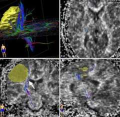
Diffusion Tensor Imaging (DTI): White matter contains the cabling (axons) connecting one part of the brain to another. In the case of the motor system, cutting the cables (corticospinal tract) will result in paralysis. This set of DTI images shows how close the corticospinal tracts (arrow, lower left image) are to the tumor (yellow). Standard MRI imaging does not show this information. UCLA neurosurgeons routinely use this information.
Photo of MRI Spectroscopy (MRS)
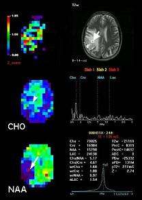
MRS: The upper right MRI shows an abnormal area in the right parietal lobe. A square "voxel" (marked by the arrow) is the area of interest within the tumor. The middle left MRS image shows the amount of choline (CHO) contained in each part of the brain. Areas of high CHO (arrow) suggest closer crowding of cells (such as a tumor). The lower left MRS shows that NAA (a chemical found in neurons, or brain cells) is low in the area of the tumor. Because tumor cells displace neurons, low NAA may also suggest tumor. In this case, a biopsy confirmed a low-grade astrocytoma.
Photo of Positron Emission Tomography (PET)
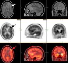
PET: The upper panel images are MRI scans showing a small low-grade astrocytoma in the left frontal lobe (white arrow). The middle panel shows a FDOPA-PET with high uptake (black arrow) in an area immediately in front of the abnormality seen on MRI. The lower panel shows the fused (overlapping) MRI and PET images. At surgery, the area identified by PET revealed the diagnostic tissue.
Photo of Image Fusion brain scan
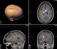
The upper left image is a three dimensional view of the head with the tumor shown in purple and the white matter tracts (identified by DTI) in yellow. The other images show the same information in three different cross sections through the brain (tumor shown by arrow).

Advanced Imaging Capabilities

Standard magnetic resonance imaging (MRI) and computed tomography (CT) imaging are important tools for diagnosing brain tumors.

These routine scans, however, do not tell us about the function of the brain surrounding the tumor. Sometimes we may desire more information about the tumor itself, such as its boundaries or whether it is actively dividing.

  • UCLA offers the most comprehensive, advanced imaging capabilities available, including very powerful 3 Tesla MRI scanners, and is a world leader in positron emission tomography (PET) scanning.

Functional MRI (fMRI)

  • Functional magnetic resonance imaging (fMRI) is an imaging technique that detects local changes in blood flow when parts of the brain are activated. For example, in order to move your fingers, the brain cells in the motor cortex become very active.

    They need more blood flow to supply oxygen and sugar to the cells. Because fMRI can detect the increase in blood flow, the imaging technique provides a picture of which part of the brain is used to move your fingers.
  • In order to detect changes in blood flow reliably, a very high-power 3 Tesla MRI unit is needed. In addition, specially trained experts are needed to interpret the fMRI data. The UCLA Brain Mapping Center has the tools and expertise to perform the highest quality fMRI imaging.
  • Functional MRI can be important for surgical planning and mapping of critical speech and motor brain regions that may be adjacent to the tumor. In special cases, further speech mapping must be done in the operating room using highly specialized techniques offered at only a few hospitals, such as Ronald Reagan UCLA Medical Center.

Diffusion Tensor Imaging (DTI)

  • Brain tumor neurosurgeons also like to know how different critical brain regions connect. Although fMRI can show where the signals begin, imaging the important connecting fibers (white matter) may be just as crucial.
  • DTI provides the surgeon with fiber-tracking information.

MRI Spectroscopy (MRS)

  • MRS is a special technique that provides some information about the chemical make-up of the tumor. MRS can help distinguish tumor from other abnormalities (such as infection).
  • MRS can sometimes identify the most malignant portion of a large tumor, guiding the surgeon to the best biopsy site.

Positron Emission Tomography (PET)

  • PET can provide unique information about tumor behavior.
  • FDG-PET assesses how much sugar (glucose) the tumor is using comparing to the normal brain.
  • FDOPA-PET looks to see if the tumor takes up DOPA, a chemical used by brain cells to make other signaling molecules.
  • PET can sometimes identify the most malignant portion of a large tumor, guiding the surgeon to an appropriate biopsy site.
  • PET can be used to follow tumors over time, assessing if they change in behavior.

Image Fusion

  • UCLA neurosurgeons use image fusion (exactly overlapping the images in three dimensional space) to bring all of the above information together in the operating room.
  • This information is used during the course of the operation to help achieve a safer, more effective surgery.
  • Intra operative image fusion. UCLA brain tumor neurosurgeons are able to bring together the advanced imaging and brain mapping techniques available at UCLA into the operating room.
  • This gives them a "road map" showing unique features of the tumor as well as the location of critical structures that must be avoided to preserve speech, walking and other functions.
Facebook Twitter Instagram Youtube LinkedIn
UCLA Health hospitals ranked best hospitals by U.S. News & World Report
  • UCLA Health
  • Find a Doctor
  • School of Medicine
  • School of Nursing
  • UCLA Campus
  • Directory
  • Newsroom
  • Subscribe
  • Patient Stories
  • Giving
  • Careers
  • Volunteer
  • International Services
  • Privacy Practices
  • Nondiscrimination
  • Billing
  • Health Plans
  • Emergency
  • Report Broken Links
  • Terms of Use
  • 1-310-825-2631
  • Maps & Directions
  • Contact Us
  • Your Feedback
  • Report Misconduct
  • Get Social
  • Sitemap