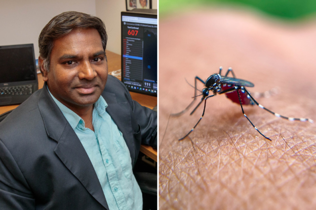
Dr. Vaithi Arumugaswami, professor of molecular and medical pharmacology at the David Geffen School of Medicine at UCLA, and Dr. Ashok Kumar, professor of ophthalmology, visual and anatomical sciences at Wayne State University School of Medicine, have received a four-year, $2.7 million grant from the National Institutes of Health to continue their study of how Zika virus damages the developing eye and to identify drug-based strategies that could protect babies’ vision.
Zika, spread mainly by Aedes aegypti mosquitoes, is best known for causing severe birth defects affecting the brain, but the virus can also harm the eyes of infants born to infected mothers. Damage to the retina and optic nerve, part of what’s known as congenital Zika syndrome, can lead to lifelong vision problems. There are currently no approved vaccines or antiviral drugs for Zika, and how the virus damages the eye remains poorly understood.
While there is no worldwide surge at the moment, the mosquito that spreads Zika virus has taken hold in parts of the United States. Locally acquired dengue cases have also been reported in the Los Angeles area, a sign that mosquito-borne viruses are becoming a concern closer to home.
“Zika outbreaks are episodic, flaring up and then fading for years, which can make the threat easy to forget,” said Arumugaswami, who’s also a member of the UCLA Broad Stem Cell Research Center and the California NanoSystems Institute at UCLA. “The time to study the virus is now, while things are quiet, not after an outbreak has taken hold and a lot of babies and families are suffering.”
In earlier work supported by a previous NIH grant, the team studied how Zika virus infects cells of the retinal pigment epithelium, a single layer of tissue that supports the eye’s light-sensing cells. Using a technique that sorts the cell’s protein complexes by size and reveals which proteins travel together, they found that Zika’s proteins appear to partner up with cell proteins that help guide eye development and manage energy. When the researchers dialed down several of these cell proteins, the virus multiplied faster, suggesting they act as natural defenders against infection. The team also screened thousands of potential drug compounds and identified several that blocked the virus and, in early tests in the eyes of infected adult mice, reduced eye damage.
The protein work was done in collaboration with Dr. Mehdi Bouhaddou and the drug screening with Dr. Robert Damoiseaux, both of UCLA.
The new funding will allow the team to examine how Zika disrupts these protein interactions at three levels: in lab-grown retinal pigment epithelium cells, in lab-grown “mini retinas” — simplified models of the developing eye grown from human stem cells —and in a newborn mouse model of Zika eye disease. Because mouse eyes finish developing after birth, this model allows researchers to investigate how infection affects a still-forming eye. Dr. Anne Zaiss, a staff scientist in the Arumugaswami lab, will support these studies.
The team will also test whether the promising compounds they’ve identified can reduce eye damage in infected newborn mice and whether they interfere with normal retinal development, a key safety consideration for any treatment that might one day be used during pregnancy.
Notably, in lab tests, these compounds also reduced infection by related mosquito-borne viruses, including dengue and West Nile, raising the possibility of broader antiviral uses.
“We need to get a drug that goes to clinical trial and benefits patients,” Arumugaswami said. “That’s the bottom line.”