A person who is having a stroke may temporarily lose the ability to speak, walk or move their arms. For many stroke survivors, however, some of these lost abilities return spontaneously during the first few weeks after the stroke. Few patients recover fully and most are left with some disability, but the majority exhibit some degree of spontaneous recovery.
Doctors and scientists don’t fully understand how this happens, because the brain does not grow new cells to replace the ones damaged by the stroke. Neurologists have generally assumed that the brain instead recruits existing neurons to take over for those that are lost.
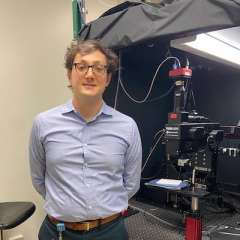
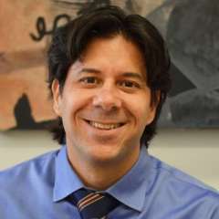
Now, new results from UCLA Health neurologists William Zeiger, MD, PhD, and Carlos Portera-Cailliau, MD, PhD, challenge that idea. In their paper published June 25 in the journal Nature Communications, they show that in mice, undamaged neurons do not change their function after a stroke to compensate for damaged ones.
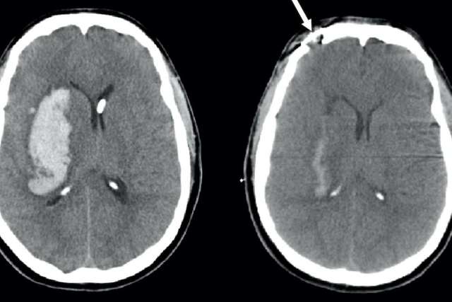
A stroke occurs when the blood supply to a certain part of the brain is interrupted, such as by a blood clot. Brain cells in that area become damaged and can no longer function. Depending on which part of the brain has been damaged, the skills lost after the stroke may include motor skills, speech or sensory perception.
Neurologists have been “mapping” the brain since the mid-1800s, when the French physician Paul Broca realized that patients who sustained damage to a certain part of the frontal lobe lost the ability to speak. Over the years, scientists have created increasingly detailed maps of which brain regions control various functions. Still, the resolution of the map has been limited by the precision of the tools available to study the living brain.
Studies in animals and humans that recorded activity across different brain regions have found that activity patterns change after a stroke, suggesting that the damaged brain can “re-map” functions from one area to another. In the last few years, innovative new tools have enabled researchers to start looking at individual neuron activation in real-time. By using a technique called two-photon fluorescence microscopy that causes neurons to light up when they are activated, researchers can observe which neurons in the animal brain are called upon during certain activities and determine if neurons that survived a stroke can assume the function of those that were lost.
“We thought, now that we have this tool where we can record the activity of neurons in the brain, we could directly test this question,” says Dr. Zeiger. By learning the functions of individual neurons, then causing a targeted stroke, the researchers could use the new technique to observe how the neighboring neurons responded.
The experiment
Mice gather information about their environment primarily through their whiskers, and each whisker transmits sensory signals to a specific group of neurons. By destroying the neurons coded to a specific whisker, the researchers could look at whether neurons for a different whisker took over for their lost neighbors.
Think of a departmental office, says Dr. Portera-Cailliau. “Imagine that several admin staff in human resources for the Department of Neurology suddenly quit their jobs one day. Instantly, the department would suffer their absence, but would try to make up for it.” The department might do this by asking HR employees from other departments to do some of the missing employees’ work on top of their own.
That’s what the re-mapping hypothesis predicts would happen in the brain. “If the hypothesis were true, we would see cells that survived the stroke start to respond to that whisker that had the stroke,” Dr. Zeiger says. “What we found was that that didn’t happen.”
Adjacent to the main column of neurons that respond to a given whisker are neurons called “surround-responsive” cells. These are located in the column for one whisker, but they react to the stimulation of a neighboring whisker. Using the office analogy, these neurons would be like employees that work on HR but happen to have their desks in a different physical office.
Logically, then, the team thought that the surround-responsive neurons would be good candidates to take over when the primary responding neurons were destroyed by the stroke.
“If the HR office suddenly shut down, you might think more people in these other offices would start doing human resource jobs,” Dr. Portera-Cailliau says. Yet not only did these other neurons fail to step up, the activity of the surround-responsive neurons themselves decreased after the stroke.
“That was pretty strong evidence against this remapping hypothesis,” Dr. Zeiger says. “It did not seem like there was bulk recruitment to take over the function which had been lost to stroke.”
More studies will be needed to gain a full understanding of what happens in the human brain after a stroke, including why spontaneous recovery happens. Dr. Zeiger says that future studies could also explore ways to induce the surviving cells to compensate for the lost neurons.
“I think it gives the field more of a direction,” Dr. Zeiger says. “Rather than assuming the brain can remap on its own, now we know that to achieve full recovery, we’re going to need a way to get cells to do things they are not already doing.”
Learn more about stroke research at UCLA Health
Caroline Seydel is the author of this article