Prostate-specific membrane antigen (PSMA) imaging provides “a more sensitive way – a better way – to detect and stage prostate cancer,” says Robert Reiter, MD, professor of urology and chief of UCLA’s Division of Urologic Oncology. This improved imaging opportunity informs treatment, “… based on what is actually happening versus what we used to know, which was probably only part of the truth.”
PSMA imaging, a type of a positron emission tomography (PET) scan highly sensitive to prostate cancer, goes beyond the already considerable limits of MRI, says Dr. Reiter, a member of the UCLA Health Jonsson Comprehensive Cancer Center. PSMA imaging enables physicians to stage patients with high-risk disease prior to deciding if the best immediate treatment strategy is surgery or radiation.
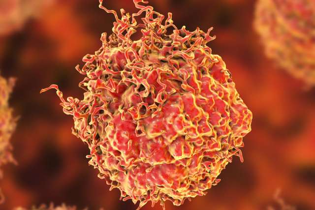
PSMA is a protein made on prostate cancer cells. This scan can detect wherever that protein is present and where there are cancer cells, Dr. Reiter says.
“It’s the most likely scan to pick up metastatic disease. And, because it is so sensitive and specific, there are very few false positives when diagnosing metastatic prostate cancer,” he explains.
In fact, PSMA allows physicians to accurately stage and detect metastatic and regional prostate cancer in up to 70% of patients with biochemical recurrence.
During PSMA testing, a clinician injects a tracer substance that can target any site and “lights up” on the PET scan. By contrast, MRI is not too useful for metastatic disease, Dr. Reiter explains. While MRI can deliver excellent images of the prostate, “PSMA PET scans actually ‘see’ the cancer and are more likely to diagnose metastatic disease,” he says. “Historically, we used CAT and bone scans, and they were very insensitive. A man had to have extremely advanced disease for us to see anything.”
Dr. Reiter’s interest in improved methods for diagnosing and staging prostate cancer began many years ago when he routinely saw men with biochemical recurrences. Their blood test cancer markers were rising, even though they already had surgery.
“We basically had no idea where the disease was. So, if we gave them radiation to the pelvis, we’d be wrong half the time,” he recalls. “Because half the time the disease would be somewhere else, like in the bone, and vice versa. Clearly PSMA imaging enables us to know the correct location to treat.”
UCLA in the lead
While PSMA imaging has been in widespread clinical use in Europe and Australia for some years, UCLA has been a leader in the use of PSMA in the U.S.
Up until a year ago, UCLA was one of only a scant few places in the U.S. doing it. Johannes Czernin, MD, chief of the Ahmanson Translational Imaging Division and a JCCC member - working with UCLA colleagues in urology, medical oncology, radiation oncology and nuclear medicine, as well as a team at UC San Francisco - spearheaded the clinical trials that resulted in FDA approval in 2022. Now PSMA imaging is being widely adopted throughout the U.S.
This broadened use of PSMA imaging is an important step forward in battling prostate cancer, the highest incidence of which is found in African American men.
Additionally, African American men have a higher risk of dying from prostate cancer in the U.S., partially based on access to care as well as the care itself.
“We are trying to provide the best care possible to benefit, and be inclusive of, all patient populations. Fortunately, PSMA imaging is covered by Medicare and most commercial insurers now, and that allows greater access,” says Dr. Reiter.
A novel approach
The benefits of PSMA imaging are being featured at UCLA. “There’s something unique we’re doing that has not been done anywhere else in the U.S.,” says Dr. Reiter. “We’re using the same imaging to guide surgery. We use the PSMA tracer the day before men with disease in their lymph nodes undergo surgery. In the operating room, we can use a gamma probe to determine where the disease is located. So, if something lights up on the scan, we can find that in the operating room. This is a novel approach.”
What’s the big take-away about PSMA imaging? It’s always about the patients, stresses Dr. Reiter. He believes PSMA imaging will positively transform the way physicians manage prostate cancer moving forward – and that could lead to better patient outcomes.