FINDINGS
Researchers evaluated motor skills and cognitive development, visual and hearing function, and brain images of children who had been exposed to the Zika virus during their mothers’ pregnancies. By the age of 12 to 18 months, significant problems were present in seven of the 112 children (6.25 percent) who were evaluated for eye abnormalities, in six of the 49 children (12.2 percent) evaluated for hearing problems, and in 11 of the 94 children (11.7 percent) evaluated for severe delays in language, motor skills and/or cognitive function who also had brain imaging.
In all, 19 of a total of 131 children (14.5 percent) had at least one of the three abnormalities.
BACKGROUND
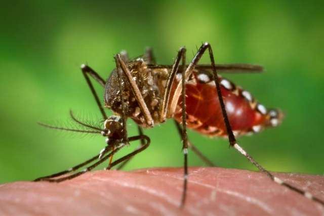
From September 2015 to June 2016, Rio de Janeiro, Brazil, experienced an epidemic of Zika virus, a then relatively unknown mosquito-borne virus that was later linked to an unusually high number of cases of microcephaly, a condition characterized by an abnormally small head, and other neurological abnormalities in infants.
METHOD
The researchers conducted at least one imaging exam (using transfontanelle cerebral ultrasound, computerized tomography or magnetic resonance imaging); an eye exam; a standard screening called Bayley III, which measures infants’ cognitive, language and motor skills; and a brainstem evoked audiometry test, which diagnoses hearing loss in newborns and young children.
IMPACT
The findings provide a reliable estimate of the frequency of neurodevelopmental, vision and hearing defects in infants and toddlers after they are exposed in utero to Zika. The research supports the use of brain imaging to help predict neurodevelopmental health in babies who have been exposed to the virus. The researchers note, however, that brain imaging failed to predict severe neurodevelopmental delays in 2 percent of cases. Also, 16 percent of infants who had abnormal brain images early in life had normal cognitive, language and motor skills by 12 to 18 months of age.
AUTHORS
The study’s authors include Dr. Karin Nielsen, professor of clinical pediatrics in the division of pediatric infectious diseases at the David Geffen School of Medicine at UCLA and UCLA Mattel Children’s Hospital; Dr. Maria Elisabeth Moreira of the Instituto Fernandes Figueira, Fiocruz, Rio de Janeiro; and Dr. Patricia Brasil, head of the Febrile Illnesses Division, Fiocruz, Rio de Janeiro.
JOURNAL
The article appears December 12 in the New England Journal of Medicine.
FUNDING
This study was supported by the National Institutes of Health’s National Institute of Allergy and Infectious Diseases and National Eye Institute, the Thrasher Research Fund, the Bill and Melinda Gates Foundation Grand Challenges Explorations, and grants from government agencies and other funders in Brazil.


