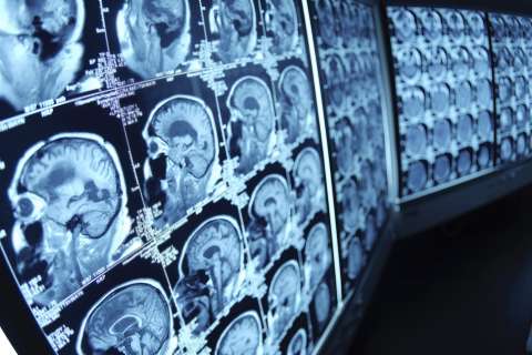
Sometimes called the “floor” of the skull, the skull base sits at the bottom of the cranial cavity, separating the brain from other facial structures such as the nose, ears and eyes. It is a delicate and complex region, surrounded by blood vessels and nerves that are essential to many bodily functions.
When a tumor infiltrates the skull base, it can cause excruciating pain, vision loss, hearing loss and hormonal issues. The best solution is to remove the tumor; however, it’s a challenging, high-risk area to operate in and around.
“Historically, these were no-go zones,” says Gregory P. Lekovic, MD, PhD, a neurosurgeon at UCLA Health and associate professor in the David Geffen School of Medicine.
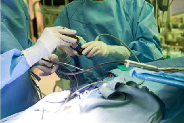
It was during the early 1990s that new endoscopic surgical techniques led to safer skull base procedures. Today, treating tumors from the bottom of the skull takes a specialized and multidisciplinary team, which can include neurosurgeons, otolaryngologists, radiation oncologists, oculoplastic surgeons, endocrinologists, ophthalmologists and radiologists.
That is what sets UCLA Health apart from other clinics and hospitals, says Marvin Bergsneider, MD, a neurosurgeon and professor at UCLA Health and director of the UCLA Pituitary and Skull Base Tumor Program. “Skull base surgery is a team sport, and I think we have the best team here at UCLA.”
Treating vestibular schwannomas (aka acoustic neuromas)
A vestibular schwannoma, previously commonly known as an “acoustic neuroma”, is a non-cancerous tumor that develops on the nerve that leads from the inner ear to the brain. Early symptoms can be easy to miss, but left untreated, the tumor can cause vertigo, balance issues, and hearing loss.
Acoustic neuroma has been, in some ways, “the highest peak” for neurosurgeons to climb, says Dr. Lekovic, who joined UCLA this past summer after a 15-year career at the prestigious House Clinic in Los Angeles, as well as serving as the director of neurosurgery research at the House Institute Foundation. Harvey Cushing, who is known as the father of modern neurological surgery, described his experience with the complicated surgery in a 100-year-old text. During Dr. Cushing’s career, the mortality rate for the removal of acoustic neuromas was about 50 percent, Dr. Lekovic says.
“Now the surgery is extremely safe. And once the medical field brought together all different specialties — neurology, ENT, radiation oncologists — it’s fascinating what arose,” he says. “It created a cross-pollination of ideas that led to better surgical outcomes and the identification of other diseases that could benefit from surgery.”
Dr. Lekovic has treated more than 1,000 patients with acoustic neuromas throughout his career.
Depending on the size and location of the tumor, the procedure typically starts with an otolaryngologist making an initial incision in the ear’s temporal bone, exposing the tumor. Then, Dr. Lekovic takes over to safely remove the tumor and preserve the patient’s hearing.
“The old way of doing things was for the neurosurgeon to make a hole in the skull and move the brain out of the way to get to the tumor,” he says. “The current skull base team approach is to safely come to the tumor first, which puts the brain less at risk.”
Dr. Lekovic’s research portfolio includes improving outcomes and techniques for acoustic neuroma surgery, as well as studying acoustic neuromas in patients with the genetic condition neurofibromatosis type 2 (also called NF2-related schwannomatosis). Dr. Lekovic says that understanding the genetic underpinnings of NF2 may give researchers a better idea of what causes the tumor in patients without NF2.
The pituitary gland and pituitary tumors: A closer look
The pituitary gland is a pea-sized endocrine gland located at the base of the brain, producing hormones that regulate growth, metabolism, and reproduction. Sometimes, small tumors (pituitary adenomas) can grow on the gland. This can lead to several serious conditions, including Cushing’s disease, which is caused by the overproduction of adrenocorticotropic hormone (ACTH) that stimulates the adrenal glands to produce the stress hormone, cortisol. Symptoms of Cushing’s include weight gain, high blood pressure, bone loss, fatigue, and diabetes.
Surgically removing the pituitary adenoma is the best option for curing Cushing's disease.
“You can try to chemically interfere with that messaging system, but there aren’t good medications for that,” Dr. Bergsneider says. “Another option is to remove the adrenal glands, but removing the adrenal glands means that a patient becomes entirely dependent on replacement cortisol. And if they miss a dose, they could die.”
Dr. Bergsneider has treated more than 1,500 pituitary tumors, along with head and neck surgeon Marilene B. Wang, MD, and endocrinologist Anthony P. Heaney, MD.
Using a minimally invasive technique that was adopted early on at UCLA, Dr. Bergsneider and Dr. Wang remove pituitary tumors through the nose with an endoscope. Tissue samples from the tumor are then sent to Dr. Heaney, who is investigating new potential medications for Cushing’s disease.
While pituitary surgery is highly safe and effective, occasionally, some patients’ tumors are so small that they are virtually invisible on an MRI scan.
“From symptoms and blood tests, we know that a patient has a tumor that is causing harmful effects, but their MRI scans come back as normal,” said Dr. Bergsneider.
This forces surgeons like Dr. Bergsneider to go into the area without a map — making incision after incision in the pituitary gland as they search for a tumor. “It’s horrible,” he says.
After nearly a decade of research, Dr. Bergsneider and colleagues at UCLA believe they have come up with a solution. They have designed a small, flexible coil that goes into the nasal cavity during surgery and allows for a signal-to-noise ratio on an MRI scanner that is 17 times higher than with a typical head coil.
“It’s like a radio — the closer you are to the gland, the stronger the signal, and the better the picture,” Dr. Bergsneider says.
The research team is now seeking approval from the U.S. Food and Drug Administration to test the new MRI coil in clinical trials.
Minimally invasive surgery through the eyelid
A different approach employed by the UCLA skull base team, which is used to remove tumors near the optic nerve and other cranial nerves responsible for eye movement and facial sensation, is called transorbital neuroendoscopic surgery (TONES).
Using this method, two surgeons work in collaboration to enter the skull through a patient’s eyelid crease. TONES provides a minimally invasive alternative for patients with multiple prior surgeries.
“We’re able to remove tumors without large incisions or craniotomy,” says UCLA neurosurgeon and associate professor Won Kim, MD. “It gives us a new corridor to enter that won’t have the same risk of wound breakdown or infection because of prior surgeries and radiation.”
In addition to taking new approaches to surgery, the success of UCLA’s skull base surgery team also lies in monthly conferences, where clinicians from across divisions come together to discuss cases and exchange ideas.
Dr. Kim says that the minimally invasive procedures and team of highly specialized experts at UCLA Health give hope to patients who otherwise thought they may have been out of options.
“We're able to bring the best surgical minds together to come up with innovative treatment plans and surgical approaches,” he says, “which allows us to operate on challenging tumors that others in the community may not have felt were operable.”
Lauren Ingeno is the author of this article.