• UCLA Health
  • myUCLAhealth
  • School of Medicine
UCLA Interventional Neuroradiology

UCLA Interventional Neuroradiology

UCLA Interventional Neuroradiology
  • Back to Main Radiology
  • About Us
    • Welcome
    • Quality Data
    • Maps and Directions
    • Research
    • In The News
    • Case of the Month
    • Fellow's Corner
    • Disclaimer
  • Diseases Treated
    • Acute Stroke
    • Arteriovenous Malformation
    • Brain Aneurysm
    • Carotid Angioplasty/Stenting
    • Cerebral Venous Sinus Thrombosis
    • Dural Arteriovenous Fistula
    • Moyamoya Disease
    • Pediatric Diseases
    • Spinal Vascular Malformation
  • Procedures Offered
    • Balloon Test Occlusion
    • CT Angiography
    • Diagnostic Angiography
    • Embolization
    • Inferior Petrosal Sinus Sampling
    • Vertebroplasty / Kyphoplasty / Sacroplasty
  • For Patients
    • Webinars
    • Patient Stories
  • Contact Us
  • Our Expert Team
  • UCLA Health
  • myUCLAhealth
  • School of Medicine

UCLA Interventional Neuroradiology

Procedures Offered

Procedures Offered

Procedures Offered

  • Balloon Test Occlusion
  • CT Angiography
  • Diagnostic Angiography
  • Embolization
  • Inferior Petrosal Sinus Sampling
  • Vertebroplasty / Kyphoplasty / Sacroplasty
  • Balloon Test Occlusion
  • CT Angiography
  • Diagnostic Angiography
  • Embolization
  • Inferior Petrosal Sinus Sampling
  • Vertebroplasty / Kyphoplasty / Sacroplasty
  1. Home
  2. UCLA Interventional Neuroradiology
  3. Procedures Offered
  4. Vertebroplasty / Kyphoplasty / Sacroplasty

Vertebroplasty / Kyphoplasty / Sacroplasty

Share this
Figure 1. A typical lumbar compression fracture (white arrow)

Figure 1. A typical lumbar compression fracture (white arrow)

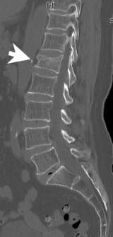
Compression fracture of the vertebral body is common. It is defined as a vertebral bone in the spine that has decreased in height by at least 15% due to the fracture (Figure 1). It is more common in the lower spine (thoracic and lumbar). Some vertebral compression fractures may heal on their own. However, in most cases, the compressed vertebrae do not fully heal since the fractured vertebral fragments continue to move even with a back brace.

Vertebral compression fractures can cause severe back pain leading to inability to perform daily activities, and potentially social isolation due to immobility. It is known, particularly in women, that vertebral compression fractures are associated with a 15% higher mortality than those without. In order to prevent this ripple effect, we can perform a minimally invasive percutaneous interventional procedure that can stabilize the fractured bone with the use of cement. Depending on the lesion, we offer 3 types of interventional procedures; vertebroplasty, kyphoplasty, and sacroplasty.

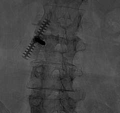
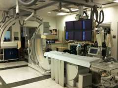
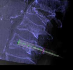
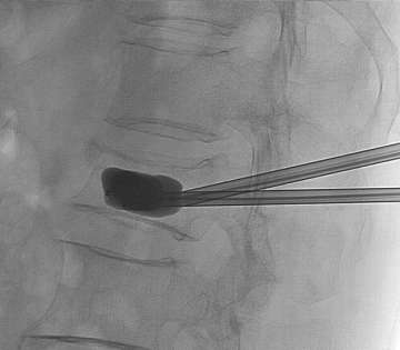
During the procedure, the patient is lightly sedated and lies on their tummy. Under x-ray guidance, we place the needle into the vertebral body (Figure 2). Unlike most pain services where a relatively simple portable C-arm X-ray machine is used, we use the state-of-the-art biplane neuro-angiosuite, which is capable of 3-D navigation in the brain and spine (Figure 3). In rare occasions, placement of the needle is challenging due to previous implantation of metal hardware that obscures the normal bony landmarks, or due to severe osteoporosis. The 3-D navigation capability provides us with the ability to very accurately place the needle without relying on the classic radiological bony landmarks (Figure 4).

Figure 2. A needle is placed into the fractured vertebral body under x-ray guidance.

Figure 2. A needle is placed into the fractured vertebral body under x-ray guidance.

Figure 3. The state-of-the-art biplane neuro-angio suite, which is capable of 3-D navigation in the brain and spine.

Figure 3. The state-of-the-art biplane neuro-angio suite, which is capable of 3-D navigation in the brain and spine.

Figure 4. A needle is being placed into the vertebral body using the state-of-the-art 3-D navigation system, enabling very precise positioning of the needle without relying on classic bony landmarks that may become fuzzy in a patient with osteoporosis.

Figure 4. A needle is being placed into the vertebral body using the state-of-the-art 3-D navigation system, enabling very precise positioning of the needle without relying on classic bony landmarks that may become fuzzy in a patient with osteoporosis.

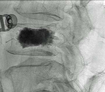
Once the needles are placed into the fractured vertebral body, cement is injected through the needles so that it can be stabilized from its inside (vertebroplasty).

If there is a need to restore the height of the fractured vertebral body, a balloon is inflated within the fractured vertebral body prior to cement injection (kyphoplasty / Figure 5). Once the bone cement hardens, it becomes an internal cast and completely stabilizes the lesion (Figure 6).

Figure 5. Balloons are inflated within the fractured vertebral body in order to regain the height and also to create a cavity

Figure 5. Balloons are inflated within the fractured vertebral body in order to regain the height and also to create a cavity into which thick cement can be injected (Kyphoplasty).

Figure 6. The cement hardens in 10-15 minutes and completely stabilizes the vertebral body from the inside.

Figure 6. The cement hardens in 10-15 minutes and completely stabilizes the vertebral body from the inside.

The patient can be discharged home the same day. In some cases, particularly after Kyphoplasty the patient may need to stay overnight. In our experience, more than 90% of the patients had complete disappearance or significant reduction of the pain over the next two weeks. For more detailed information, please feel free to contact us.

Like Us on Facebook Follow Us on Twitter Subscribe to Our Videos on YouTube Follow us on Instagram Connect with Us on LinkedIn Follow us on Pinterest
UCLA Health hospitals ranked best hospitals by U.S. News & World Report
  • UCLA Health
  • Find a Doctor
  • School of Medicine
  • School of Nursing
  • UCLA Campus
  • Directory
  • Newsroom
  • Subscribe
  • Patient Stories
  • Giving
  • Careers
  • Volunteer
  • International Services
  • Privacy Practices
  • Nondiscrimination
  • Billing
  • Health Plans
  • Emergency
  • Report Broken Links
  • Terms of Use
  • 1-310-825-2631
  • Maps & Directions
  • Contact Us
  • Your Feedback
  • Report Misconduct
  • Get Social
  • Sitemap
Like Us on Facebook Follow Us on Twitter Subscribe to Our Videos on YouTube Follow us on Instagram Connect with Us on LinkedIn Follow us on Pinterest

Sign in to myUCLAhealth

Learn more about myUCLAhealth