Understanding & Surviving a Brain Attack
Find Your Care
Radiologists are experts in all types of imaging, including advanced techniques.
Call today to find medical imaging near you and schedule your imaging procedure (MR, CT, PET, Dexa, Ultrasound).
Stroke: the "Brain Attack"
A stroke is an interruption of the blood supply to any part of the brain; it is sometimes called a "brain attack." If blood flow is stopped for longer than a few seconds and the brain cannot get blood and oxygen, the brain cells can die causing permanent damage. Other names for stroke are cerebro- vascular disease, CVA, Cerebral infarction, and cerebral hemorrhage.

"I was sitting at my desk at work when I had the stroke and suddenly I could not say a single word or move my right side. But, thanks to the pioneering work of the Interventional Neuroradiologists and the Stroke Team, I had a miraculous recovery. I returned to work in just a month and am able to enjoy all of my favorite outdoor activities just like always." ~ Nancy Richardson ~
There are 2 types of strokes: Ischemic and hemorrhagic strokes
Ischemic stroke is when a blood vessel carrying blood to the brain is blocked by a blood clot. Hemorrhagic stroke is when a blood vessel breaks open, causing blood to leak into the brain.
Stroke is the 3rd leading cause of death and the leading cause of nursing home admission.
Acute interventions can reduce the severity of the brain damage if the patient is rushed to the hospital the moment a stroke occurs.
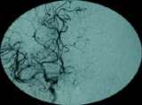
fig. 1: BEFORE the procedure: an angiograph showing the connecting vessels of the right to the left side at the base of the brain only allowed some flow into the first portion of the left middle cerebral artery, but the remainder was blocked.
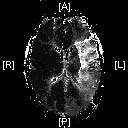
fig. 2: BEFORE the procedure: a perfusion MRI showing the left middle cerebral artery territory, the white area is where the blockage occur.
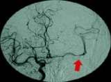
fig. 3: AFTER the procedure: an angiograph shows the reopening of the middle cerebral artery.
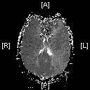
fig. 4: AFTER the procedure: a perfusion MRI showing the normalization of flow to the left middle cerebral artery territory.
Symptoms of Stroke
For Nancy Richardson, she experienced neck pain and headaches 3 days prior to her acute stroke where her right-sided arm, leg, and face became paralyzed and she had difficulty speaking. In Richardson's case, the major artery on the left side of her brain spontaneously developed a tear in the wall that resulted in blood clot formation which obstructed the internal carotid artery that impacted Richardson's areas of strength, sensation and speech.
A perfusion MRI study was conducted and determined the area of the impaired blood flow, figure 2 shows the left middle cerebral artery territory, the white area is where the blockage occur; here, the connecting vessels of the right to the left side at the base of the brain only allowed some flow into the first portion of the left middle cerebral artery (fig. 1).
Treatment of Stroke
Richardson was given rTPA intravenously to help the body break down the blood clot. A small catheter from the femoral artery was simultaneously placed in the groin up through the blocked internal carotid artery and into and beyond the blood clot on the left middle cerebral artery were more rTPA was infused and was successful in reopening the majority of the middle cerebral artery (fig. 3).
A second perfusion MRI was completed after treatment and showed normalization of flow to the effected area (fig. 4).
Richardson was able to speak & move her right side the night after the procedure. She returned to work and recovered remarkably.
Quick Links
- Stroke Symptoms & Treatments
- Interventional Neuroradiology
- MERCI Retriever
- Patient Referral
- Our Locations