Pathogenesis of Ear Diseases Laboratory
Pathogenesis of Ear Diseases Laboratory - UCLA Head and Neck Surgery
Introduction
Our mission is to study important ear diseases at the cell and molecular level. A primary focus of research of this laboratory is the pathogenesis of otitis media, including bacteria-host cell interactions, cell signaling, host-innate immune responses, mechanisms of the inner ear inflammatory response to otitis media pathogens, and the development of an innovative strategy to treat or prevent otitis media using antimicrobial innate immune molecules. Another area of research focuses on the elucidation of the molecular and cellular mechanisms involved in inner ear homeostasis, particularly in regard to the structure/functions of the endolymphatic sac, and the pathogenesis of Meniere's disease and endolymphatic hydrops.
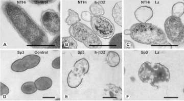
Ultrastructural evidence of killing of OM pathogens (nontypeable H. influenzae and S. pneumoniae) when treated with antimicrobial innate immune molecules such as human ? defensin 2 (h-?D2) and lysozyme (Lz).
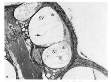
(A) Middle turn of cochlea of the chinchilla 12 hours following endotoxin instillation. Inflammatory cell infiltration and bleeding (*) are seen in perilymphatic spaces and spiral ligament with mild endolymphatic hydrops (arrow). SV: scala vestibuli, ST: scala tympani.
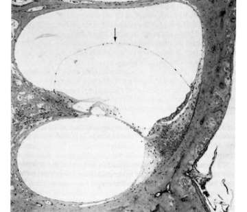
(B) Endolymphatic hydrops (arrow) are seen in middle turn of chinchilla cochlea four weeks following endotoxin instillation into the bulla. No cellular infiltration is seen in the perilymphatic space.
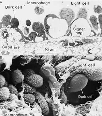
Ultrastructure of endolymphatic sac epithelial cells (chinchilla). Transmission electron microscopic view showing dark and light epithelial cells. Exaggerated intercellular space forming "signet ring" indicates active fluid transport by the epithelial cells (A). Scanning electron microscopic view showing light and dark cells and microphages (B). Light cells are characterized by abundant microvilli, in contrast to dark cells with very few or no microvilli.
Current Research Projects
- NIH R01DC005025: Innate Immunity in Otitis Media Pathogenesis
- NIH R33DC011862: Role of Spiral Ligament Fibrocytes in Immune-Mediated Inner Ear Damage
Members
- Director: David J. Lim, MD
- Co-Director: Sung K. Moon, MD, PhD
- Staff Research Associate: Jeong-Im Woo, PhD
- Staff Research Associate: Sung-Hee Kil, PhD
- Laboratory Technician: Yoo Jin Lee, BS
Recent Publications
Patent
- U.S. Patent No. 6,207,157 (3/2001): Conjugate Vaccine for nontypeable H. influenzae
- U.S. Patent No. 6,358,688 (4/19/2002): Immortalized human middle ear epithelial cell lines
- U.S. Patent No.7,338,936 B2 (3/4/08): Use of Antimicrobial Proteins and Peptides for the Treatment of Otitis Media and Paranasal Sinusitis
- U.S. Patent Pending: Cell Line Derived from the Epithelial Lining of the Human Endolymphatic Sac in the Inner Ear
Contact Information
David J. Lim, MD
[email protected]
(213) 353-7021 (office)