Radiology
How to Perform an MRI Guided Breast Biopsy
By Ethan Zaccagnino MD, and James Chalfant MD
The following guide is meant to serve as a general overview of an MRI guided breast biopsy. Depending on institutional preferences and equipment used, there may be mild variations in the steps for the procedure.
Step 1: Pre-Procedure
- Discuss the risks, benefits, and alternatives to the procedure with the patient
- Obtain written and verbal consent from the patient
- MRI technologist brings the patient to the MRI suite
Step 2: Imaging
- Technologist will gently compress the breast using a breast biopsy coil with grid localizer in place
- Radiologist confirms appropriate compression of breast tissue and performs adjustments if necessary
- Obtain images of the breast pre-contrast administration (fig 1) (sequences used can vary depending on institutional preferences)
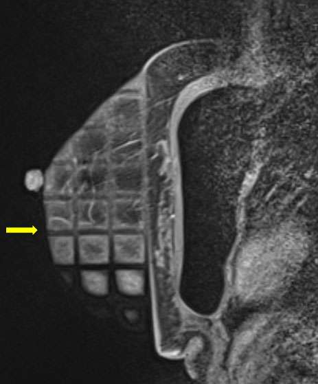
Figure 1. Sagittal T1-weighted fat saturated pre contrast images demonstrate a biopsy grid overlying the lateral breast in compression. In this patient with silicone implants, the implant must be excluded during compression.
- Radiologist and technologist perform additional adjustments of breast compression, if necessary, followed by repeat pre-contrast imaging
- Administer MRI contrast and perform post-contrast imaging
- Identify the biopsy site and determine lesion location using the grid localizer and depth within the breast from the grid (fig 2). Depending on institution, biopsy targeting software and/or post-processed images (such as subtracted images or reformatted images) may be used.
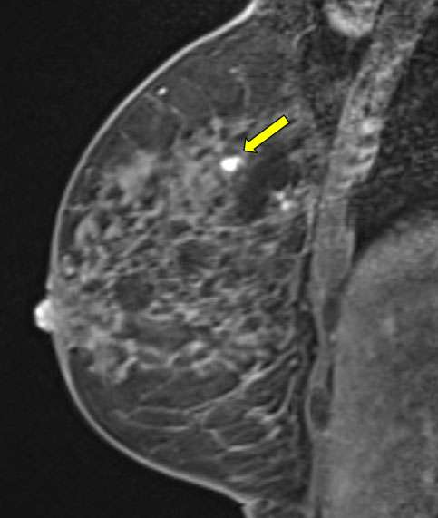
Figure 2. Sagittal T1-weighted fat saturated post contrast images demonstrate the biopsy target, a focus of enhancement.
Step 3: Procedure Preparation
- Prepare the biopsy site in the usual sterile fashion
- Drape the area with sterile drapes
- Achieve local anesthesia using a combination of lidocaine and lidocaine with epinephrine
Step 4: Biopsy
- Advance a trocar and sheath through a skin nick to the biopsy site
- Replace the trocar with an obturator
- Confirm accurate placement with a repeat scan
- Perform biopsy with vacuum-assisted biopsy device to obtain multiple specimens
Step 5: Post-Biopsy
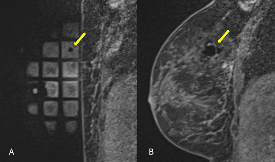
Figure 3A and 3B. Sagittal T1-weighted fat saturated post contrast images demonstrate the biopsy tract and post sampling changes.
- Confirm biopsy of the target site with a post-biopsy scan (fig 3a and 3b)
- Place a micro-clip
- Achieve hemostasis with manual compression of the biopsy site
- Send the specimens for histological analysis
Step 6: Post-Procedure
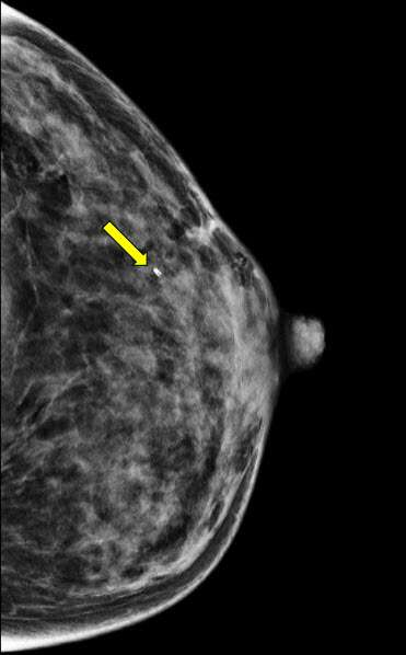
Figure 4. Post-biopsy craniocaudal mammogram confirms post-biopsy micro-clip placement.
- Perform a post-procedure mammogram to demonstrate accurate placement of the micro-clip (fig 4)
- Provide the patient with written and verbal aftercare instructions
References:
- Adrada BE, Guirguis MS, Hoang T, Spak DA, Rauch GM, Moseley TW. MRI-guided Breast Biopsy Case-based Review: Essential Techniques and Approaches to Challenging Cases. Radiographics. 2022 Mar-Apr;42(2):E46-E47. DOI: 10.1148/rg.210126. Epub 2022 Feb 4. PMID: 35119965; PMCID: PMC8906341.
- Niketa C, Pang KA, Lim JW. Challenges in MRI-Guided Breast Biopsy and Some Suggested Strategies: Case Based Review. Diagnostics (Basel). 2022 Aug 16;12(8):1985. DOI: 10.3390/diagnostics12081985. PMID: 36010334; PMCID: PMC9407085.
- Papalouka V, Kilburn-Toppin F, Gaskarth M, Gilbert F. MRI-guided Breast Biopsy: A Review of Technique, Indications, and Radiological-pathological Correlations. Clin Radiol. 2018 Oct;73(10):908.e17-908.e25. DOI: 10.1016/j.crad.2018.05.029. Epub 2018 Jul 2. PMID: 30041954.