A new imaging technique approved by the U.S. Food and Drug Administration (FDA) for use at UCLA and UC San Francisco is a significant advance over current standard-of-care imaging to detect prostate-cancer cells that have migrated beyond the prostate.
Known as prostate-specific membrane antigen PET imaging, or PSMA PET, the technique offers greater precision to localize newly diagnosed high-risk prostate cancers or prostate cancers that recur after treatment, say UCLA Health physicians who have used the technique to diagnose and stage the disease in more than 3,500 patients over the last four years. Their research with UCSF colleagues led to the approval for use at the two sites.
Among men in the U.S., prostate cancer is the second most common cancer and the second-leading cause of cancer death.
“Because the PSMA PET scan has proven to be more effective in locating these tumors, it should become the new standard of care for men who have prostate cancer, for initial staging or localization of recurrence,” says UCLA Health nuclear-medicine specialist Jeremie Calais, MD.
When prostate cancer is initially diagnosed, or when rising prostate-specific antigen (PSA) levels indicate it has recurred after treatment, knowing if and where the tumor cells have spread beyond the prostate is critical to treatment decisions, as well as to ensuring the precision of the treatment. Because prostate-cancer treatments often come with significant side effects, greater accuracy in the so-called staging process can prevent overtreatment, as well as potentially improving the efficacy of therapy by pinpointing the location of cells that might otherwise be missed.
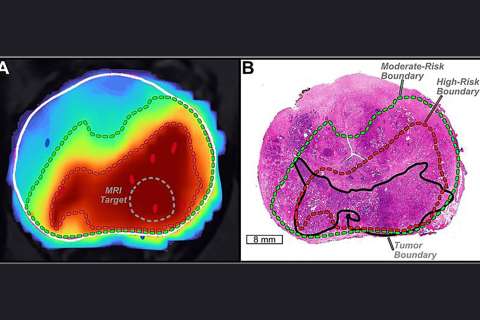
PSMA PET uses a radioactive tracer drug called 68Ga-PSMA-11, which, once injected into the patient’s body, attaches to prostate-specific membrane antigens — proteins known to be overexpressed in prostate-cancer cells. Using PET imaging, physicians can then see where the cancer cells are located based on the signal coming from the metabolism of the drug.

In a clinical trial, the UCLA research team compared PSMA PET — which has been used successfully in countries outside of the U.S. for a number of years — with fluciclovine PET, the current standard of care for prostate-cancer imaging. They found that PSMA PET was able to detect significantly more prostate lesions in men who had undergone prostate-cancer surgery but had experienced a recurrence of their cancer, as well as identifying cancer cells that would otherwise have been missed by the standard imaging approach.
Leaders of the UCLA Health and UCSF teams that helped to make PSMA PET available to U.S. patients for the first time hope that the technique will be made more widely available as an imaging test for men with high-risk or recurrent prostate cancer. The FDA approval currently limits use of PSMA PET to the UCLA and UCSF manufacturing methods, and reimbursement from Medicare and private insurers is not yet established.

Nonetheless, demand for PSMA PET is high. Johannes Czernin, MD, chief of UCLA Health’s Ahmanson Translational Theranostics Division, notes that patients are being referred from all over the country, and that there is a waiting list for the procedure.
“This major effort between the UCLA and UCSF nuclear medicine divisions, urology, oncology, radiation oncology and radiology will significantly change for the better how this cancer is detected and treated,” Dr. Czernin says.
To learn more about PSMA Pet imaging at UCLA Health, go to, uclahealth.org/psma
Dan Gordon is the author of this article.