CT: Computed Tomography
Find Your Care
Radiologists are experts in all types of imaging, including advanced techniques.
Call today to find medical imaging near you and schedule your imaging procedure (MR, CT, PET, Dexa, Ultrasound).
CT scan was first introduced in the 1970's. CT scans excel at defining bone and vascular problems such as complex fractures and narrowing or aneurysms of blood vessels. CT scans provide greater clarity and reveal more details than regular x-ray exams.
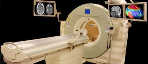
Using specialized equipment and expertise to create and interpret CT scans of the body, radiologists can more easily diagnose problems such as cancers, cardiovascular disease, infectious disease, appendicitis, trauma and musculoskeletal disorders.
ACR Gold Standard of Accreditation
CT is commonly used to detect tumors and monitor treatment response. CT angiography on new state of the art scanners is able to show the coronary arteries without ever placing a catheter in the arteries! CT can be used to create internal endoscopic views of the colon & lungs.

Patient Information (PDFs)
- CT Procedure Screening Questionnaire - English
- CT Procedure Screening Questionnaire - Spanish
- Prep for Adult CT Virtual Colonography
- Prep for Adult CT Virtual Colonography with Barium
Quick Links
- Your Heart Imaged Gently: Coronary CTA with Lowest Practical Dose
- Dual Energy CT Angiography Enhances Diagnostic Capabilities
- SOMATOM Definition: Faster Than Every Beating Heart
- Our Locations
- Patient Referrals
Related Links
- Radiology Info: Computed Tomography (CT or CAT Scan)
- Radiology Info: Safety in X-ray Examinations
- Image Gently
- Image Wisely
RADIATION SAFETY STATEMENT:
Your physician has determined that a Computed Tomography (CT) scan is necessary in the course of your health care. CT scans are an essential tool in the practice of medicine for diagnosis of disease as well as for the assessment of response to treatment. The detailed assessment of anatomy and function that CT imaging provides requires the use of X-rays. X-rays are a form electromagnetic radiation similar to visible light but much higher in energy. We are constantly exposed to natural radiation in the course of our daily lives including terrestrial (e.g. radon), cosmic, and radiation from within our own bodies. The additional radiation from a CT exam varies considerably depending on the type of exam performed. but is typically on the order of what a U.S. citizen receives from natural sources in 1-5 years. The additional risk to an individual from medical X-ray is not well known but believed to be very small. However, as the risk is assumed to not be zero and is believed to increase with radiation dose it is important that optimal quality images are obtained at the lowest X-ray dose possible. At UCLA, we are constantly evaluating our CT imaging protocols with teams of academic and clinical experts - including specialty radiologists, licensed technologists, and board certified medical physicists - to ensure that the lowest possible radiation dose is used to achieve the diagnostic image quality required for the study.