Palpable Lump
by Randy Chang, MD, Lucy Chow, MD, Fadi Dahoud, and Bo Li, MD
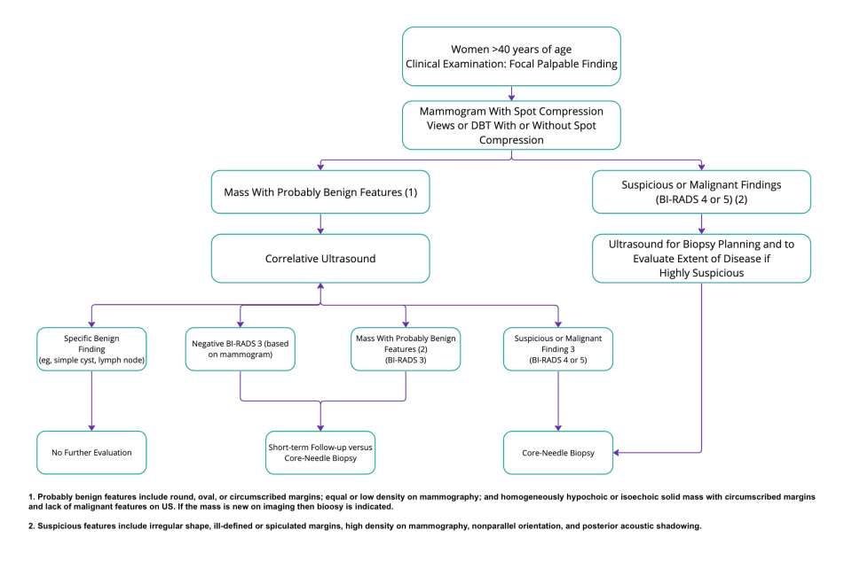
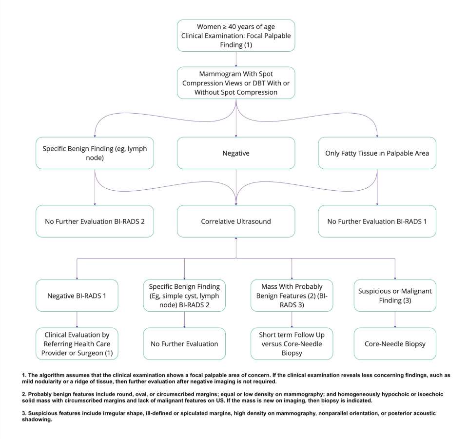
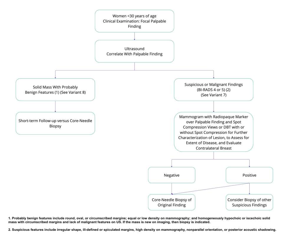
Palpable breast lumps can cause anxiety for patients. Most palpable breast lumps are benign. However, a new palpable breast mass is also a common presenting sign of breast cancer. In almost all cases, imaging evaluation is necessary to characterize the palpable lesion. Recommended imaging options include diagnostic mammography and targeted breast ultrasound (US). The choice of modality is dependent on patient age and degree of radiologic suspicion. Any highly suspicious mass detected by imaging or palpation should undergo core biopsy with image guidance (US, stereotactic, tomosynthesis, or MRI) unless contraindicated (1, 2).
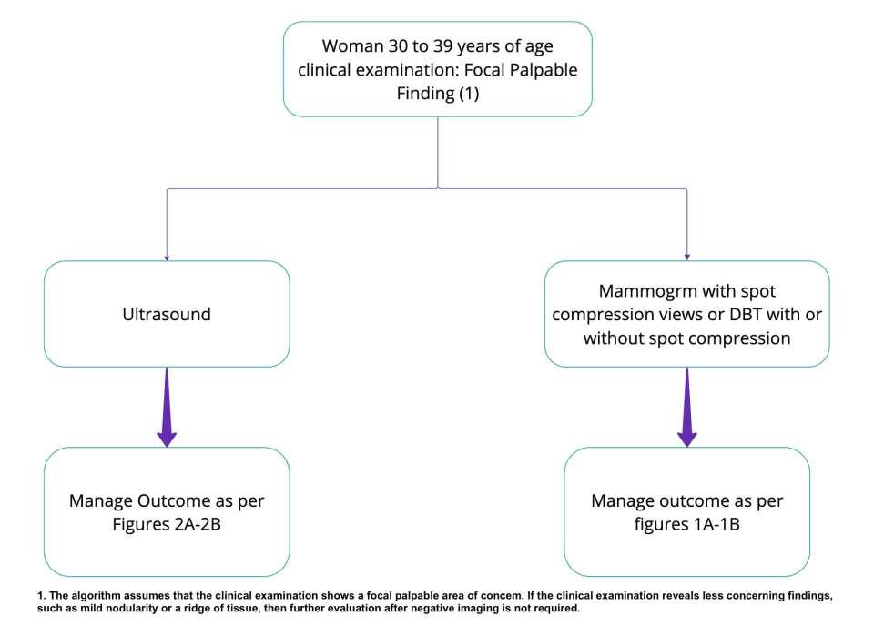
Diagnostic mammography (DM) is indicated for women ≥ 30 years of age who present with a palpable lump. If a clearly benign correlate for a palpable finding can be identified on DM, no further imaging may be indicated with appropriate clinical follow-up. If an imaging correlate is identified that is not clearly benign or the DM is negative, targeted US should be performed. Additionally, digital breast tomosynthesis (DBT) can address some of the limitations encountered with standard DM views. DM or DBT may also be helpful for women ages 30 to 39 with palpable lumps and women younger than 30 with palpable lumps and suspicious findings on US (1, 2). Further details can be seen in Figure 1A-B and 3.
Mammography is also generally safe for pregnant and lactating women. Radiation is focused on the breasts, and a lead shield is placed over the belly for further protection. It is also recommended for lactating women to breastfeed or “pump and dump” prior to mammography (3, 4).
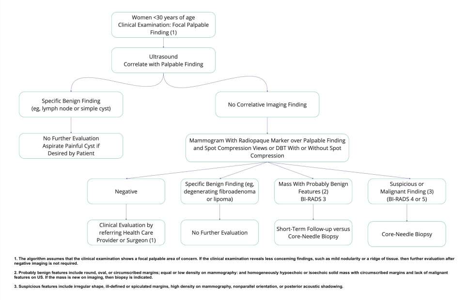
Ultrasound is the first-line recommendation for a palpable lump in women under 30 years of age. If there is a suspicious finding on US, DM or DBT is recommended for further characterization to identify features of malignancy. US is also an essential next step in evaluating women ≥ 30 years of age with a palpable mass and either a negative mammogram or a finding not clearly benign on mammogram (1, 2). Further details can be seen in Figure 2A-B.
References
- Expert Panel on Breast Imaging:, Moy L, Heller SL, Bailey L, D'Orsi C, DiFlorio RM, Green ED, Holbrook AI, Lee SJ, Lourenco AP, Mainiero MB, Sepulveda KA, Slanetz PJ, Trikha S, Yepes MM, Newell MS. ACR Appropriateness Criteria®Palpable Breast Masses. J Am Coll Radiol. 2017 May;14(5S):S203-S224. DOI: 10.1016/j.jacr.2017.02.033. PMID: 28473077.
- Daly C, Puckett Y. Approach New Breast Mass. [Updated 2020 Aug 2]. In: StatPearls [Internet]. Treasure Island (FL): StatPearls Publishing; 2021 Jan-. Available from: https://www.ncbi.nlm.nih.gov/books/NBK560757/
- Robbins J, Jeffries D, Roubidoux M, Helvie M. “Accuracy of diagnostic mammography and breast ultrasound during pregnancy and lactation.” AJR Am J Roentgenol. 2011 Mar;196(3):716-22. Erratum in: AJR Am J Roentgenol. 2011 May;196(5):1237. DOI: 10.2214/AJR.09.3662. PMID: 21343518.
- Expert Panel on Breast Imaging:, diFlorio-Alexander RM, Slanetz PJ, Moy L, Baron P, Didwania AD, Heller SL, Holbrook AI, Lewin AA, Lourenco AP, Mehta TS, Niell BL, Stuckey AR, Tuscano DS, Vincoff NS, Weinstein SP, Newell MS. “ACR Appropriateness Criteria® Breast Imaging of Pregnant and Lactating Women.” J Am Coll Radiol. 2018 Nov;15(11S):S263-S275. DOI: 10.1016/j.jacr.2018.09.013. PMID: 30392595.