Pulmonary Angiography and Embolization
Find your care
Our board-certified physicians diagnose and treat many vascular conditions. Call 310-481-7545 to learn more about interventional radiology services.
Treatment for:
Pulmonary AVMs (arteriovenous malformations)
Why it’s done:
Pulmonary AVMs act as direct conduits between the pulmonary artery and the pulmonary vein, which reduces blood oxygen levels, and also allows clots and bacteria to bypass the normal filtration process of the lung capillaries. Pulmonary AVMs can also rupture, causing serious bleeding. Embolization of pulmonary AVMs greatly reduce these risks.
How it’s done:
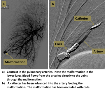
An interventional radiologist uses X-rays to guide a small catheter from the femoral vein at the groin and into the pulmonary arteries. Contrast injection is performed to locate the pulmonary AVMs. Each AVM is then accessed using a catheter or microcatheter, and blocked with small fibered coils or plugs.
Level of anesthesia:
Conscious sedation.
Risks:
Small risks of bleeding, infection, stroke, and coil dislodgment. Contrast dye and X-rays are used.
Post-procedure:
Four hours of lying flat in recovery; then discharge home the same day. Some pain with coughing or deep breathing is normal and should resolve in several days.
Follow-up:
A follow-up CT is usually performed 1-3 months after embolization to confirm complete blockage of the pulmonary AVMs. CT is then repeated approximately every 5 years to check for re-opening of the AVMs or growth of new AVMs.
For More Information:
For more information or to schedule an appointment with one of our IR physicians, please call 310-481-7545.