Benign Disease (Infertility)
Find your care
Our radiologists lead the way in prostate imaging. We offer the newest techniques to better detect and stage prostate cancer. Call 310-481-7545 to find out more about prostate imaging and treatment options.
History
- Young man presents with infertility (azospermia)
- Workup and medical history otherwise noncontributory
- Muellerian duct remnant in central upper prostate may obstruct ejaculatory ducts
- Left seminal vesicles are atrophic
Imaging
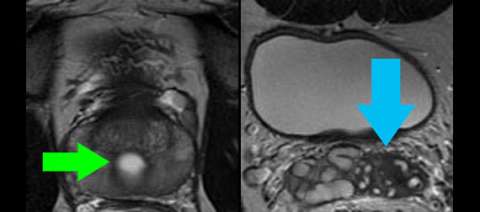
LEFT: Coronal high-resolution T2-weighted image shows a Muellerian duct remnant cyst in the superior prostate (green arrow) and atrophic seminal vesicles on one side only (blue arrow). A utricle cyst, which is much more common, would like lower in the prostate at the level of the verumontanum.
RIGHT: Sagittal reformat of isovolumetric T2-weighted image shows the location of the Muellerian duct remnant cyst (green arrow) with respect to the urethra and bladder.
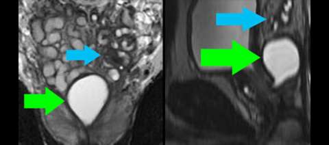
LEFT: Axial cross-section through the lower part of the Muellerian duct remnant cyst (green arrow) shows it lies midline in the peripheral gland. The prostate itself is normal.
RIGHT: Axial cross-section above the level of the prostate shows the seminal vesicles are atrophic on one side only (blue arrow) although the other side appears somewhat disordered, likely related to obstruction from the cyst (not pictured at this level).